|
APTENIA CORDIFOLIA
A SHORT ILLUSTRATED MONOGRAPH
part 2: reproductive organs
Walter
Dioni
Durango (Dgo.) Mexico
|
|
Macrophotographs
were taken with a
Canon A300 digital camera, with a resolution of 1024 x 768 pxs. and
trimmed or
reduced to fit them to this article. The photomicrographs were taken
from sections
made with a mesotome,
using my National Optical
microscope, with planachromatic optics and an integrated digital camera
of 0.4
Mpx, at 640 x 480 pxs. Backgrounds were cleaned, pictures were trimmed,
or
mosaics were made to reconstruct organs, using PhotoPaint. I believe
that the
illustrations on the two articles of this illustrated monograph of Aptenia
cordifolia, clearly shows the utility that the mesotome can have in
fast researches
on certain plants and, mainly, in
teaching botany.
Key words: Aizoacea, Aptenia cordifolia,
mesotome,
teaching botany
|
|
THE FLOWER
Aptenia N.E. Brown, is a genus
pertaining to
the Aizoacea Family.
Aptenia cordifolia (LF)
Schwante, the better known species, has a long history. It was first
described
by the son of Carolus Linnaeus,
the so-called Father of
Systematics
that dominates the scene of the classification of living beings
still today,
in spite of important new concepts that probably will impose a more
modern
version to supplant it.
For those who need
to refresh on the basic elements of
the taxonomic categories, references were given in the first
article on this species. As it is usual, different authors
interpret
some facts in a different form, and that causes some species to be
been
assigned successively to several genera. The following one is a list of
the
known names for Aptenia cordifolia.
The
letters between parentheses
(L.f.) refers to the first author who described the species and
means Linnaeus
filius, son of Lineo, also known as
Carolus Linnaeus. In his time he also was an important botanist,
eclipsed by
the enormous importance of his father.
Aptenia cordifolia (L.f.)
Schwantes
Synonyms
Litocarpus
cordifolia (L.f.) Bowl.L.
Tetracoilanthus
cordifolius (L.f.) Rappa
&
Camaronne
Mesembryanthemum
cordifolium
L.f.
A
scientific description of the flower of Aptenia can
be read in
HTTP://www.efloras.org/florataxon.aspx?flora_id=1&taxon_id=102363
But, as we did with
the vegetative organs, I prefer to
present the facts in graphical form.
Magnoliopsida (which we knew
before as Dicotiledoneae) are the most evolved plants. Like the
mammals,
which are the end of the evolution of animals, the magnoliopsida
also tried
successfully to protect their embryos, and their development, in
special closed
organs. Although, of course, with structural differences that
responds to
different ecological necessities, because plants are live entities
fixed
to the
ground and developed reproductive methods to fit to that fact.
The strategies of Aptenia,
then, are the following
ones:
The plant grows
thanks to the development of buttons
at the end of their stems which give origin to new stems, leaves or
flowers. In Aptenia
it is easy to recognize the buttons which we will call "foliar" (Lb)
from
those that we will denominate "floral" (Fb)
|
|
|
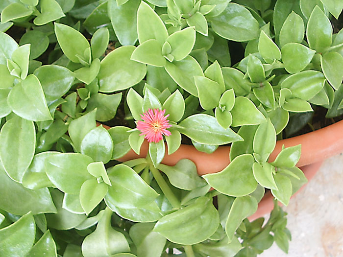
Fig. 1 – the plant, (click to see the
labeled photo).
|
|
|
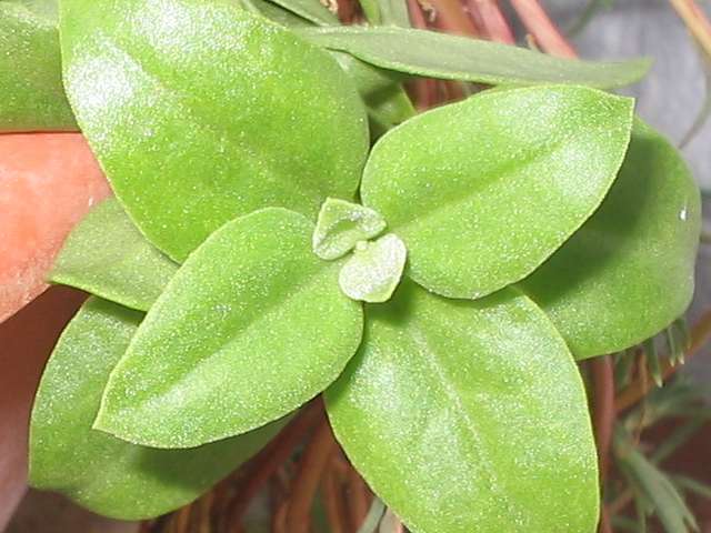
Fig. 2 – Foliar button from which only
stems and leaves are developed.
|
|
|
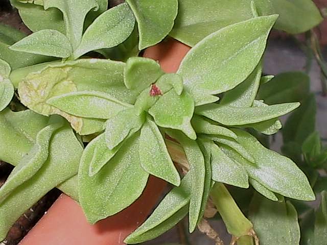
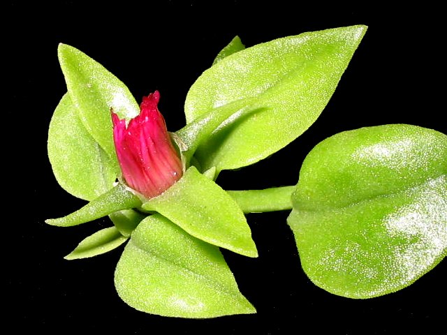
Fig. 3 - the floral button is
distinguished because the sepals (that is to say the modified leaves
that
will
form the calyx) are different: two are more or less similar to
normal
leaves, but the other two have the form of a horn or claw, thin and
curved. The
beginning of a handful of red petals appears in the junction of the
sepals.
|
|
|
|
Fig. 4 - the flower develops directly,
and it opens with the light of the day.
|
|
|
Fig. 5 - At night the flower is closed.
(Note: as a reference to this characteristic, Mesembrianthemum,
the genus to which the plant was originally ascribed, means "midday flower".)
|
|
|
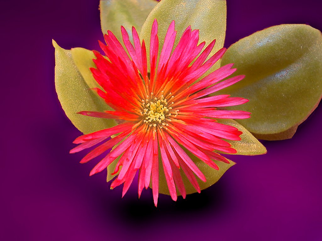
Fig. 6 - My plants generate two types of
flowers: sterile ones, (see fig.4) without visible stamens; the
others
are complete and fertile. By its symmetry this is an actinomorphic
flower and because it has all the sexual organs (masculine and
feminine) it is
denominated a perfect flower. By counting, assigned
to the
flower are 140 elements of the corolla including petals and staminodes
(see
picture
7).
It is a weird thing that in my plants (and
those of some other gardens in Cancún) only approximately 1 of
each 5 or 6
flowers is a perfect flower, the others show only petals and staminodes.
|
|
|
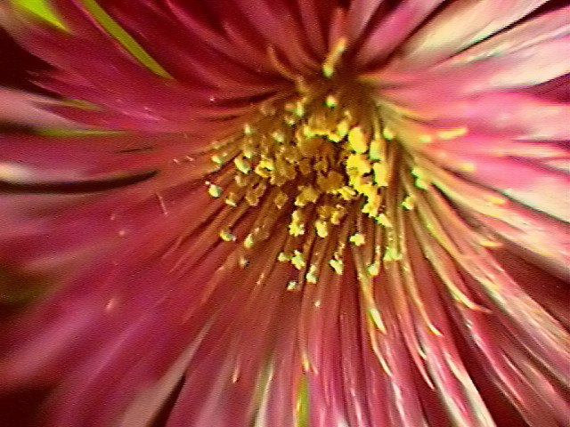
Fig. 7 - (Click to see the labeled picture.) The corolla has many layers of petals (Pt) in
whose interior several circles of stamens
with yellow anthers (St) are seen. Outside the normal
stamens there are some circles of stamens without anthers (they only
show the
filament) that are denominated staminodes (ET). This photo was
taken
using the “Köhler microscope" previously described, with
the
Obj. X 4.
|
|
|
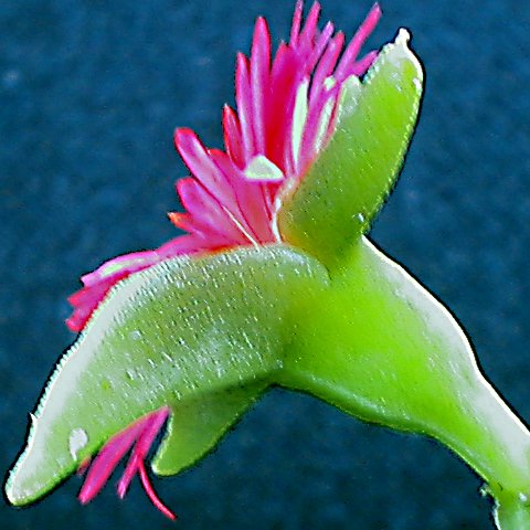
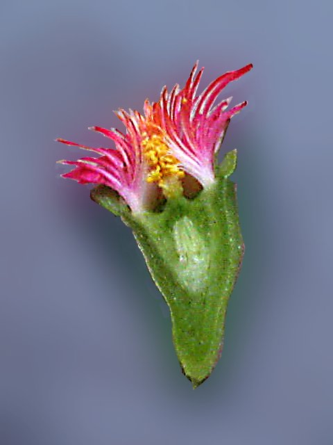
Fig. 8 - a lateral view of the flower shows
that the 4 sepals over the petals (together,
sepals and petals form the corolla) fuse to form the
more or less conical receptacle or
hypanthium
in which the feminine organs or carpels lodge (a carpel is the
set of
the ovary, style and stigma). In this flower the carpel
practically does
not have a "style", (see figs. 9 and 12).
|
|
|
Fig. 9 - a longitudinal section of the
immature flower shows in more detail the relations of all these
elements. (Click
to see the labeled image), petals, Pt; stamens, St; stigma, st; 2
carpels, c1 and c2, are shown in the section; ovules, ov; and receptacle, R). The set
of photographed
elements constitutes what it is denominated the perianth.
Petals and sepals were trimmed to allow
a better dissection. It's evident, of course, that the section has left
out the
pedicel or short stem on which the flower develops.
|
|
|
|
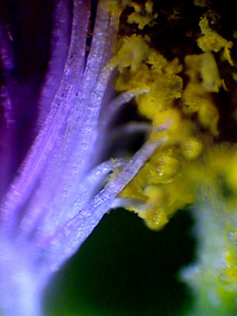
Fig. 10 - The stamens join together at
its base (obj. X 40, il. top light).
|
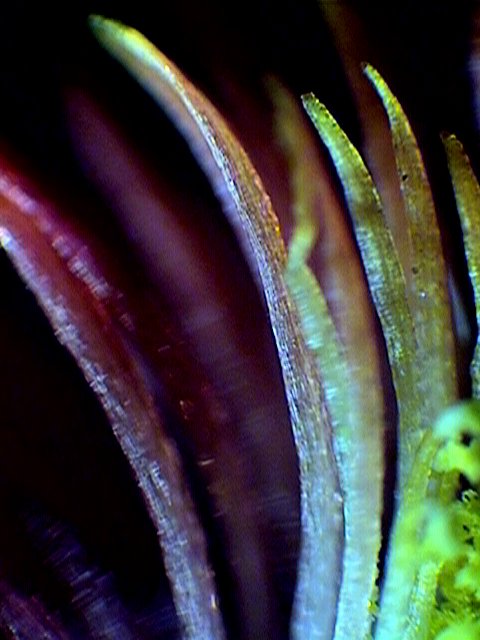
Fig. 11 - the staminodes, has only the
filament, without the anther.
|
|
|
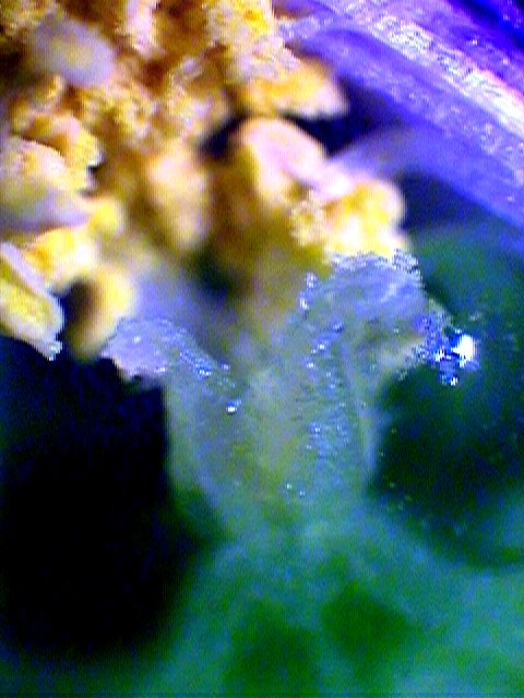
Fig. 12 – the stigmas are practically seated
at the ovary’s end, without a style.
|
|
|
|
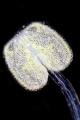
Fig. 13 - the stamen filament has a package
of nourishing vessels, and the anthers are developed in its end, as a
double sac.
Obj. X 10, Darkfield.
|
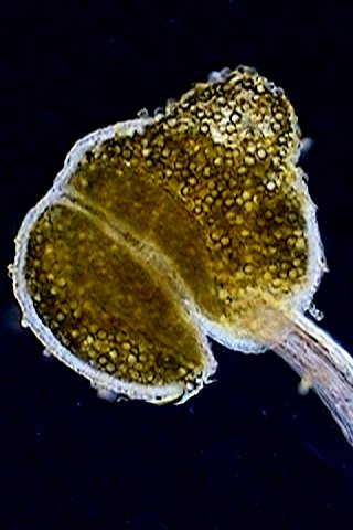
Fig. 14 – on maturing anthers develops
the pollen grains, where
the male cells are lodged. Normally, once mature, the anthers in this
flower
open by one longitudinal dehiscence furrow. This anther is not
yet mature
as to open itself, the pressure of the coverslip has broken its
envelope. Obj.
X10, Darkfield.
|
|
|
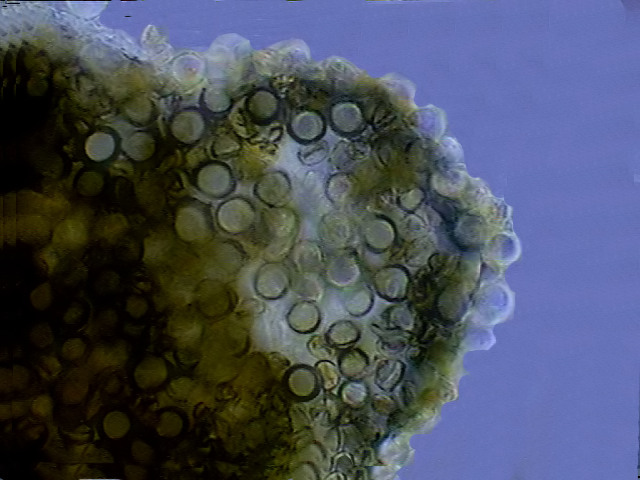
Fig. 15 - the pollen grains seen with
the Obj. 40x, brightfield.
|
|
|
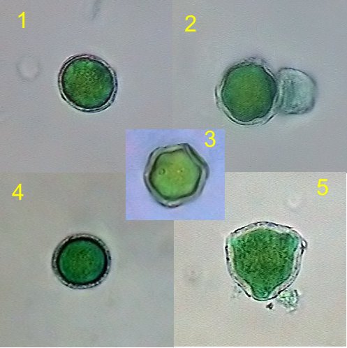
Fig. 16 - several types of pollen grains
present in the same slide. Obj. 100x HI, Stained with Fast Green,
mounted in
Glycerin Jelly.
|
|
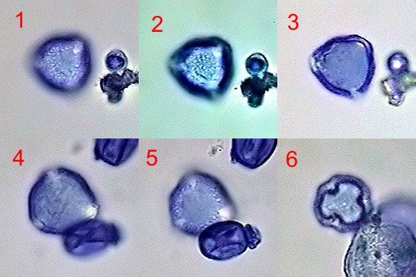
To better study the
small pollen grains
of this flower (the larger grains measured 24 micrometers (fig 16.4)
and
the
smaller 18, (fig.16.5) I used two different methods. First I fixed the
materials
in 70% alcohol, stained them with an aqueous solution of FastGreen and
mounted
in glycerin jelly.
I was surprised
that the grains were of
irregular size and forms. The probably viable pollen grains were only a
small
proportion of the total.
Carlberla
developed a staining and
fixing solution for the fast examination of pollen samples whose
original
formula is:
Glycerin.
|
5 ml
|
| Ethyl alcohol (95%). |
10 ml |
| Water |
15 ml |
| Sat.
sol. of fuchsine. |
1 drop. |
Not having some of
the elements I used a
modification:
| Glycerin |
20 ml |
| Methyl alcohol |
40 ml |
| Water |
60 ml |
| Gentian violet (1%
aqueous solution) |
1 drop |
The stronger
staining power of the
gentian violet demanded the dilution.
|
|
|
Fig. 17 - 1, 2, 3, three planes of the
same pollen grain. 4, 5, two planes of another one. 6, a very different
geometry. Obj. X 100, Immersion, Brightfield.
Results were the same, although the punctuated
nature of the exine could be better observed. Proportionally only a few
pollen grains
showed a normal shape. As seen in fig . 16 (2 and 5)
and in this mosaic, the viable pollen grains are tricolpate. ( Many
thanks to Andre Advocat from the French Forum Microscopies for
the determination.)
|
|
|
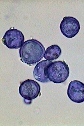
Fig. 18 - most of the pollen grains (taken
from 3 different flowers) were irregular.
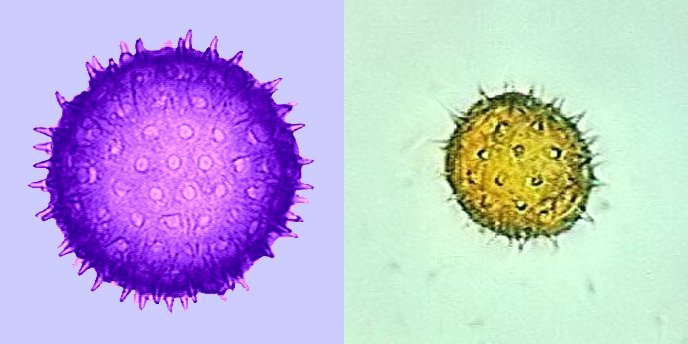
Thinking that the applied treatment (staining-fixing
solution) could distort the results, I applied the same technique to
pollen of Morning
Glory, Ipomea purpurea, and also to
the one of a small wild composite flower, whose species I don’t know.
In both
cases all the numerous grains gathered had not only a similar diameter
but also
identical morphology.
|
|
|
Fig. 19 – At left, the pollen of Ipomea
purpurea, a little squashed by the
coverslip. At right the pollen of a small wild composite, of unknown
species. Both with the 40 x Obj.
|
|
Adding this
fact to the low incidence of
fertile flowers, it occurred to me, without any certainty, because I am
not a
trained botanist, that this may be due to the fact, reported in the
following
article on the Internet, and elsewhere, that plants known as the red
variety
of Aptenia cordifolia, can be really a
hybrid cultivar.
HTTP://www.mediterraneangardensociety.org/plants/Aptenia.cordifolia.cfm
The most commonly grown plant, usually grown
under the cultivar name of 'Red Apple', is considered by some
botanists
to actually be a hybrid between Aptenia cordifolia and the
closely
related Platythyra (Aptenia) haeckeliana. It certainly shows
the vigor
common among bi-generic hybrids, carpeting large areas easily in a season or two.
|
|
|
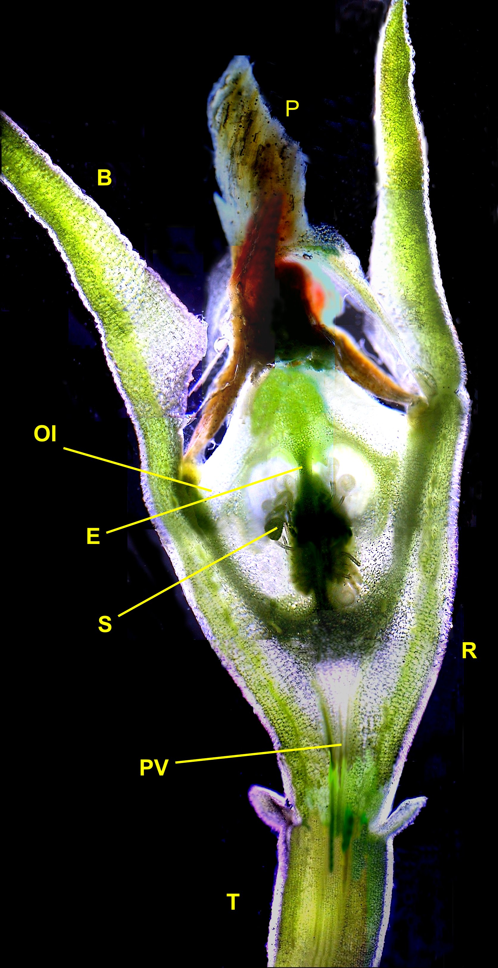
Fig. 20 - Once fertilized the flower
closes its petals, which will dry and fall, and start to produce the
fruit.
This longitudinal section of a young fruit is a mosaic reconstructed
with 8
independent images. (Click the picture if you want to see a larger
image.) T - pedicel; PV -
Vascular package which feeds the carpels; R - receptacle, that
contains the carpels;
P - dry petals still adhered to the young fruit; B - sepals; OI -
inferior
ovary, located underneath the corolla. The flowers that have this
disposition are
denominated epigynous (Epi, over, ginos, the gineceum or
set of
feminine reproductive organs.) As it can be seen, the ovary is
"submerged" in the receptacle or hypanthium. In the varied (and
complicated, according to the author) botanical terminology, this is
also denominated pericarpel, whereas the petals that normally
are
interpreted as
"the flower" are over that. Obj. X 4. Darkfield Illumination
Note: carpel is
the set of ovary + style + stigma. As it can be seen in figure 22 this
flower
has 4 fused carpels.
|
|
|
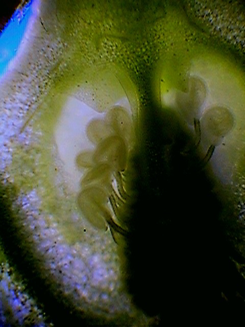
Fig. 21 - as it is seen in the previous
image and in this longitudinal section, through two carpels, the ovules
of each
ovary are adhered to a central axis and lodged within a cavity
denominated the locule.
|
|
|
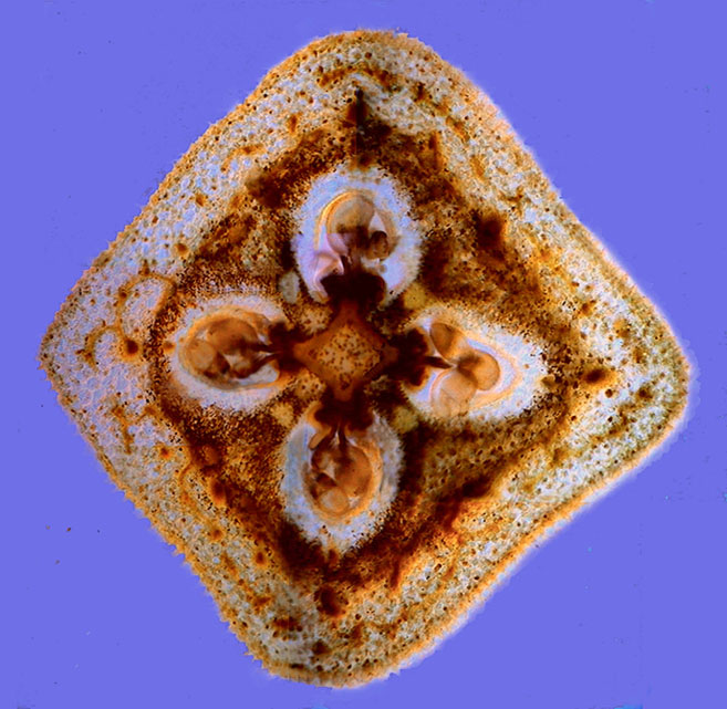
Fig. 22 - this cross section at half length
of the hypanthium shows that they have four locules, each one
corresponding to a carpel. The anatomical elements of
the ovary are
better identified in the following image. Because of problems due to
illumination and color variations when the section moves over the
microscope
stage to produce the images needed for the mosaic, this one required 11
different pictures. The original image was 1316 x 1282 pxs. Obj. X 4,
Rheinberg Illumination
|
|
|
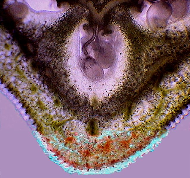
Fig. 23 - Each future seed is bound to
the axis by a filament or funiculus, adhered by its base to a placenta,
and with the future seed in the other end.
(Click to see a labeled picture.) E - central
Axis; PC placentae; F - funiculus; Ov - ovules; Sp - septum separating
two
locules; End.- endocarp, inner wall of the locule; Pr - pericarp or
wall of the
ovary; Ec - Ectocarp, external covering of the ovary; Hz - beams or
vascular packages
that feeds the pericarp. Mosaic of two images.
|
|
|
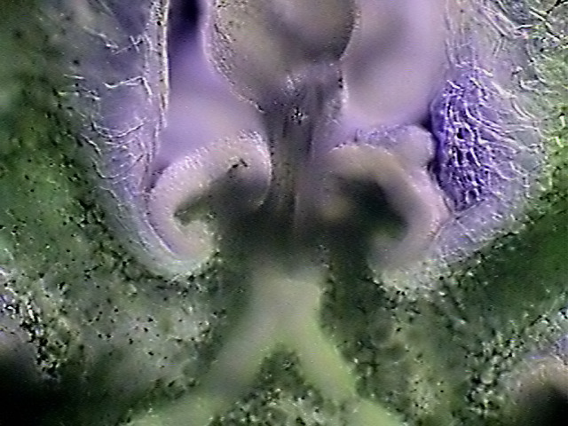
Fig. 24 - the zone of insertion of the
funiculus in the placenta. X40, Darkfield.
|

|
|
Fig. 25 – a locule cross-section. As it
were seen in the 17 picture, the ovules are adhered over the whole
length of
the axis. This style of placentation
is named axial placentation. Mosaic of two images.
|
|
|
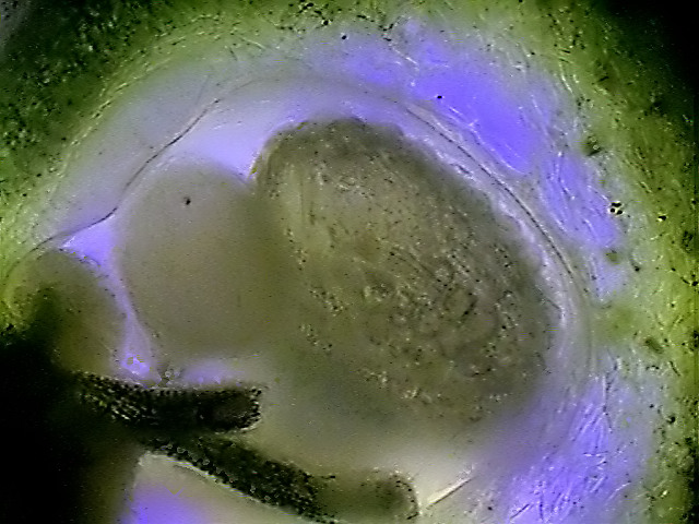
Fig. 26 - the mature seed accumulates
food around the embryo and protects the total adding a hard outer
layer,
provided with superficial tubercles. In this figure an almost mature
seed
can be
seen.
|
|
Mature carpels
produce the fruit. In the
consulted electronic bibliography the fruits of Aptenia
are named capsules, a fruit type common in the Aizoacea.
But the appearance of the fruits of
my
plants (see Figs. 28 and 29) are more similar to a "pome", a type of fruit in which the
pericarp
thickens, becomes succulent, and holds the seeds in a reduced central
space. It
is interesting that the best example of this last type of fruits is the
apple.
|
|
|
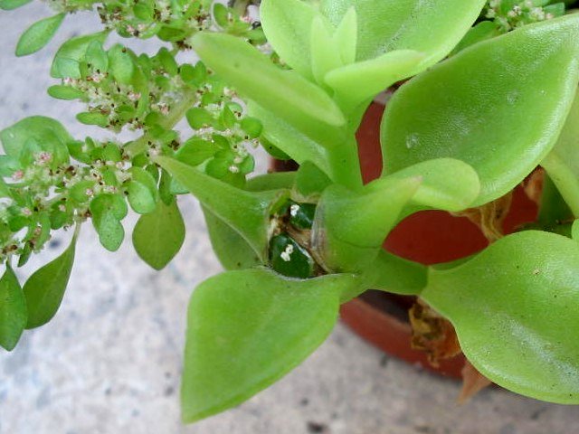
Fig. 27 - aspect of the mature fruit in
the plant.
|
|
|
|
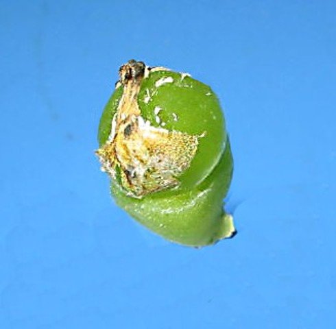
Fig. 28 - fruit with the rest of the sepals
and petals peeled off. There are seen the 4 stigmas corresponding to
the
4
carpels.
|
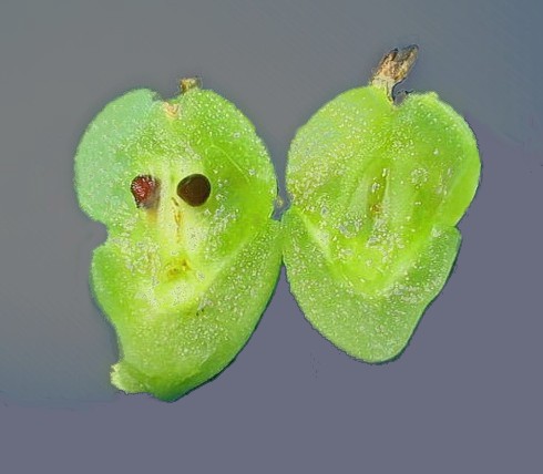
Fig. 29 - longitudinal section of the fruit, still
lodged in the receptacle. |
|
|
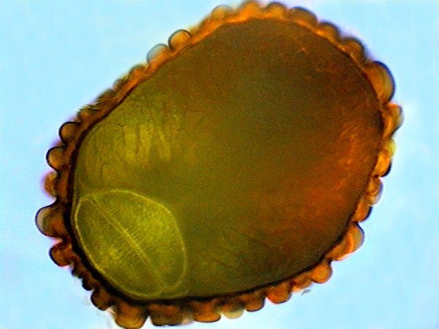
Fig. 30 - a seed cut by the mesotome,
showing in its contour the characteristic superficial tubercles.
|
| The future plant is sleeping in the seed waiting
for suitable conditions to develop and grow. Nevertheless, although
in an earlier
article it was said that Aptenia reproduction
is by seeds, in fact gardeners actually use only cuttings (the seeds
are
very small
and would be difficult to gather). The plants crawling stems root
themselves to
the immediate ground, which gives it a high invading power. The
importance of
the seeds in the reproduction of the plant in the wild would have to
be better
investigated. |
Comments to the author,
Walter
Dioni , are welcomed.
© Microscopy UK or
their
contributors.
Published in the May 2005 edition of
Micscape.
Please report
any
Web problems or offer general comments
to the Micscape
Editor.
Micscape is the on-line
monthly
magazine of the Microscopy
UK web
site at Microscopy-UK
©
Onview.net Ltd, Microscopy-UK, and all contributors 1995 onwards. All
rights
reserved. Main site is at www.microscopy-uk.org.uk with full mirror at www.microscopy-uk.net.
|