In a basic biology course with a lab, almost every student gets to look at an amoeba under a microscope. If it’s a prepared slide with preserved specimens, then it is almost certainly highly unexciting or, to put it another way, dead boring. To see a living specimen can, however, be quite a different matter, especially if it is active. Observing the protoplasmic flow, the constant shape-shifting, and the engulfing of prey can be a quite fascinating experience. However, then it’s on to the next organism, quite possibly a Paramecium, for after all, what is an amoeba but a drop of jelly that flows around a bit.
Well, as it turns out amoebae are much more than first meets the eye and they occur in surprising morphological variety and display a wide variety of intriguing behaviors. In a basic teaching lab, the standard species is Amoeba proteus. Oddly enough, this species is relatively uncommon in the wild. Decades ago, a biological supply company discovered that A. proteus was easy to culture in large numbers and the cultures turned out to be stable so, they began to market them extensively to high schools, colleges, and universities. This was fine for a basic survey course but, of course, A. proteus is no more an ideal model for the types of Rhizopoda than Paramecium is for the Ciliata. So, in this album/essay, I want to give you a glimpse into the extraordinary realm of amoebae. There is, of course, a major disadvantage to what I am presenting, because it is all static and so, we’ll try to work around that partially by using a few computer tricks to bring out some detail that we might otherwise miss.
Naturally, we shall begin with A. proteus. I was fortunate enough a few years back to find some specimens in a high lake–about 8,000 feet–and I kept them in culture for several years. If you haven’t seen them alive, it’s worth trying to find them or buying a small culture. Remember to look at them with a stereo scope in addition to your compound scope. If you are lucky, you’ll encounter specimens moving up from below toward you, thus reminding you that these are not flat 2-dimensional creatures, but indeed actively 3-dimensional ones. First, let’s look at a brightfield image.

Next, an image taken with Nomarski Differential Interference Contrast (DIC) which indeed,because of the contrast, allows us to see more detail.

I then took an image and applied the computer graphics “invert” function combined with pseudo-darkfield and here is the result.

In these images, you can see the macronucleus, vacuoles, and protoplasmic ridges. A. proteus is a stocky, robust sort of organism and will engulf most anything in its path that seems to be a juicy morsel. However, when it comes to the issue of a voracious appetite, it doesn’t hold the record by any means.
I was observing some small soil amoebae and I will give you 3 examples of what I regards as Herculean gastronomic efforts. First off, let’s look at a little guy who is apparently a devoted vegetarian.

The strand of filamentous algae is more than twice as long as the amoeba’s present extension.
If we invert the image, we get a different view and see quite clearly the cell wall demarcations in the algal filament.

However, if you’re a carnivore or an opportunistic omnivore, you might, in spite of your size, tackle a big juicy rotifer who happens along. In this instance, we have a slightly larger amoeba modifying a strategy from the Old West of “circling the wagons” to defend against invading Indians; but, in this case, the amoeba is circling the rotifer to keep it from escaping and ultimately to digest it. So, what’s amazing is that this little creature has wrapped itself around this rotifer to create a vacuole wherein it will proceed to digest it. The first image was taken with Normarksi DIC.

I then inverted the image and added a black background for contrast.

Next, I altered the color contrast and shifted to a white background.

These 3 images provide slightly different perspectives and, from my point of view, slightly different revelations of detail as well.
Next, we encounter a little critter who is truly ambitious and struggling to “pull himself together” as it were. He also is trying the “encirclement” strategy. He is tackling a Euplotes, a hypotrich which has fused ciliary bundles called cirri and it is a strong swimmer. As a consequence of the enlarged vacuole on the upper left of the Euplotes, I suspect that it may be incapacitated due to pressure from the cover glass. As you will see in the images, the amoeba has just made a start at its attempt to engulf the Euplotes. I’ll show you 2 images, the first is a modified brightfield image using color with a black background.

The second is the original brightfield image.

These are examples of the so-called “naked amoebae”. The small amoebae are soil organisms and show considerable variety and there are a relatively large number of species. The taxonomy of these forms is a nightmare and it takes a specialist to navigate the labryinth of minute morphological distinctions. There are, however, rather distinctive forms which have gained notoriety as documented or potential human pathogens. Naegleria fowleri when it gets into the sinus cavities through the nose can invade the brain and produce an extreme form of meningitis which is nearly always fatal. Valkamphia and a few other forms are also suspected pathogens. However, not all amoebae are naked and I want to turn now to some extraordinary examples of “shelled” amoebae. Later on, we shall return to another amazing example of a “naked” amoeba.
A common testate amoeba is Arcella vulgaris found in fresh water ponds and mosses. Its test of shell is entirely organic and I like to call it the “donut” amoeba.

Seen from the side, the test is roughly hemispherical. It should be noted, however, that in different species, the shape of the test can be rather different and even have spiny processes.
There are groups of amoeboid organisms that can be regarded as, in one sense, transitional between the “naked” amoebae and those that have calcareous or siliceous tests. Clearly, Arcella is one such, but there are others such as, the Heliozoa and Gromia. Heliozoans or “sun animalcules” have rod- or needle-like axopodia which extend out beyond the central spherical mass of the organism. These are supported by microtubules and one can observe protoplasmic flow along these structures. I’ll show you 3 images. The first is brightfield with altered color background.

The second image is the image above with graphic inversion and color background alteration.

The third is a monochromatic image which reveals good detail.

Next step up, we’ll consider Gromia, which taxonomically inhabits a kind of nether world and some years back, I devoted an article to Gromia which is a bewildering beastie. Here, I’ll simply show you an image and provide a very brief description.

In some says, it rather strongly resembles the foraminifera (hereafter, forams), but it has an organic test and so is regarded as being essentially a testate amoeba. It can project pseudopodia out multi-directionally, which are five, six, seven times, or even more, its body length. Protoplasm streams along these in both directions–wonderful to watch!
Since I mentioned forams, let’s move on to a consideration of a few forms of these creatures that build calcareous tests, i.e., they are basically composed of chalk. Many of the fossilized forms have undergone mineral replacement and so might seem quite different from the tests of living forams. Some have even undergone the process of pyritization and produced dazzling metallic tests. Most forams are microscopic, but 2 forms reached a distinctly macroscopic size, the fusulinds and nummilites. Both types have been found that achieved a dimension of 2 inches or slightly larger, particularly with nummilites which tend to be roughly circular and flattened. Fusulinids, on the other had, are tubular in shape, rather like a capsule with tapering ends. Cross sections of these can be highly pleasing aesthetically.

And here is the same image using the invert graphics function.

We can see all the little chambers and the marvelous Fibonacci spiral.
Forams occur in a staggering variety and have been intensively studied because up until relatively recent times deep sea core samples were examined for forams as indicators of oil deposits. New technologies have largely replaced this methodology, but the extraordinary beauty and diversity of forams still merits intensive attention and careful examination.
Because these tests or shells are opaque, or at least semi-opaque, I experimented with a technique using various kinds of oils to enhance the visibility of both internal and surface detail.
More recently I have been using certain bits of computer graphics magic as well.
Here is a typical and quite lovely form.

The inverted image is interesting in that it gives a slightly different view of surface structure. Sometimes the invert function can draw our attention to internal structure in such a way that we gain a better sense of the morphology of the test.

Consider the example below: first, a brightfield image and then an inverted image.


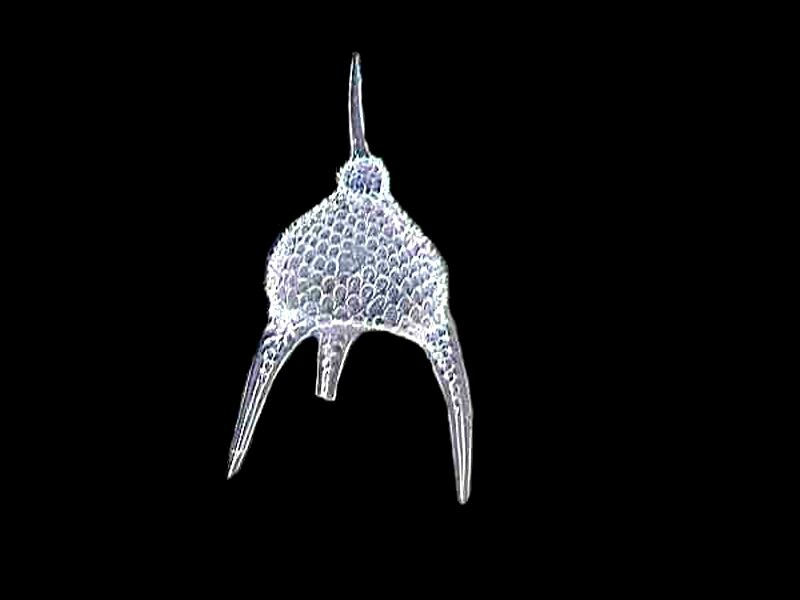
Radiolaria, although related to foraminifera, are very different in a number of crucial respects. First of all, and fundamentally, radiolarian tests are largely composed of silica, that is, glass. There is another closely, related group, the Acantharians, which have tests composed of Strontium sulfate. The diversity of these two groups is astonishing and mind-bogglingly elegant. The specimens I have are quite lovely, but don’t rival the really elaborate forms to which I will provide a couple of links later on. We’ll start simply.
One afternoon, I was sorting through some wonderful samples of forams given to me by a colleague who was a micro-paleontologist. After a couple of minutes, I realized that in addition to forams, there were a lot of radiolarian tests. These were samples from the Andaman Sea off the coast of Burma (now Myanmar). Right off, my attention was caught by some tiny, white, spiny spheres and they were numerous enough that I allowed myself to go into vandalism mode and, using some of my best micro-tools, split a number of these spheres to try to get a peek inside. Unfortunately, no genies appeared, so I was unable to make any wishes, but what I did find was quite intriguing.
First let’s take a look at the lovely, little spiny sphere.

When I split it open, I was amazed and delighted to discover a series of internal struts emanating from a tiny central core. I took a closeup and used a pseudo-darkfield background.

And then, a monochromatic image on a white background.

I would guess–without any evidence–that a strut connects to each of the spiny knobs on the surface. It makes elegant sense to me, even though it may be completely wrong.
For quite some time, almost all of the radiolaria samples available in this country came from Barbados. A couple of biological supply houses used to provide small tubes of concentrated specimens which were excellent material and cost about $15 to $20 a tube, but even these relatively small samples contained thousands of radiolarian tests. Now, unfortunately, such samples are no longer available and two biological supply houses now offer only prepared strew slides.
Some of the tests look, to my eye, rather like satellites or space capsules. For example:

or, a nice monochrome image

which I like even better with the invert function and a black background.

This might be the creation of a highly trained artist’s etching on a piece of black scratch board. In any case, I find it most attractive.
Next up, moon landers.

And again an inversion version.

Or imagine a planet made out of glass, again in 2 views, monochrome and inverted.


Politicians, these days, are always prattling on about transparency; well, they wouldn’t last long on a planet like this.
And then there are those forms that immediately strike me as alien or belonging to some different dimensions. I’ll give you 4 examples here.


The last 2 are of special interest. As I mentioned before, virtually all of the specimens commercially available to amateurs are from Barbados where there are some massive deposits. However, radiolaria are ubiquitous and I got to wondering about what some of the Pacific Ocean forms look like. So, I wrote to a West Coast specialist in radiolaria and he kindly sent me half a dozen slides of Pacific forms, two of which you see in these last 2 images.
As promised earlier, I am now going to provide you with 2 links to some of the most wonderfully elegant forms of radiolaria and acantheria which rival the beauty of almost anything on the planet (link1, link 2).
Sometimes it’s hard to grasp how all of these different and diverse forms are legitimately characterized as amoebae. I’m about to make that even harder. I told you earlier that we would return to another remarkable type of a naked amoeba, in this case, Thecamoeba which I dubbed the Shar-Pei of the amoeba-world. You can find out why here.
Thecamoebae are voracious algae eaters and seem to have a prediliction for filamentous forms. This first image is a living specimen which has ingested some filaments which I stained with Neutral Red which in highly diluted solutions is virtually non-toxic. This image was taken with Nomarski DIC. Note that the large vacuole on the left seems to be convex.

When the image is inverted, the vacuole appears to be concave and the filaments take on a light-bluish cast.

However, if you’re not fond of the color purple, then we can modify the background with the following result.

Finally, sometimes Thecamoebae manage to roll filaments into coils. Or, it’s possible that the coils are already formed in the external environment and amoebae simply ingest them in that form. That, however, seems somewhat unlikely to me as I have not come across such coils of this type of filamentous algae in the surrounding environs of the Thecamoebae. The first image is Nomarski DIC and you can see the coils clearly.

The final image is an inversion of the image above.

Ordinarily I’m not a great fan of pink, but in this image, it is, I think, quite effective. And a last item of interest is that the concave vacuole in the Nomarski image is still concave in the inverted image and I don’t know how to explain that. However, as I have grown older, I have encountered more and more things that I can’t explain.
I hope you’ve gotten a bit of enjoyment out of this brief foray into the world of amoebae.
All comments to the author Richard Howey are welcomed.
Editor's note: Visit Richard Howey's new website at http://rhowey.googlepages.com/home where he plans to share aspects of his wide interests.
Microscopy UK Front
Page
Micscape
Magazine
Article
Library
© Microscopy UK or their contributors.
Published in the March 2020 edition of Micscape Magazine.
Please report any Web problems or offer general comments to the Micscape Editor .
Micscape is the on-line monthly magazine of the Microscopy UK website at Microscopy-UK .
©
Onview.net Ltd, Microscopy-UK, and all contributors 1995
onwards. All rights reserved.
Main site is at
www.microscopy-uk.org.uk .