Close-up View of Deadnettle Hybrid 'Purple Dragon'


A Close-up View of the

Deadnettle Hybrid 'Purple Dragon'


Lamium maculatum (hybrid)


by Brian Johnston   (Canada)


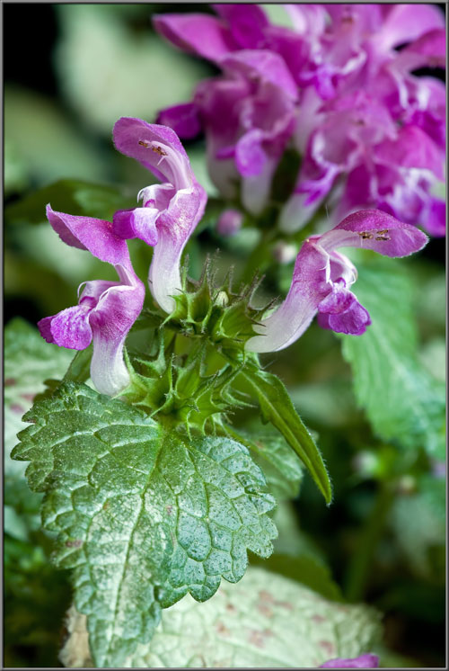
This colourful, and very attractive plant, grows to a maximum height of about 20 centimetres and is a perfect example of the phrase “Good things come in small packages”.  Its many deep purple-red flowers contrast beautifully with the silvery-green leaves.  In suitable climates, the plant is an evergreen;  unfortunately, the climate where I live is an extremely unsuitable one!

Leaves are dark green around the edge, silvery in the interior, and have purplish spots randomly sprinkled over the surface.  It may be for this reason that one of its common names is the spotted deadnettle.


Groups of buds grow from the leaf axils, and the example shown is at a very early stage of development (about 3 mm in diameter).


When a group grows a bit larger, it becomes possible to see the five spiky sepals surrounding each bud.  No colour is apparent at this stage.


Eventually, each bud appears as a small white sphere nestled in a depression, and surrounded by the five, pointed lobes of the fused sepals.


In the image below, there are two rings of sepals, the upper one, with buds, and the lower without.  After blooming the entire corolla and reproductive structures fall away, leaving the developing seeds at the base of the sepal depression.


Closer views of  the spherical buds reveal that they are completely covered by very fine hairs that make the bud look furry.


Now the buds begin to take on a pink colouration.  Notice the tiny colourless insect in the second image.


Conventional wisdom suggests that the ‘beauty’ of a plant is centred in its flowers.  Although this is undoubtedly true, the bud stage sometimes gives the flowers a good run at overthrowing that wisdom.  If you look carefully, in some of the images you can detect the unfurled ‘hood’ that will eventually become the tall, overhanging petal in the final blooming flower.  Note that at this stage, the bud has finally grown out beyond the spiky tips of the sepals.


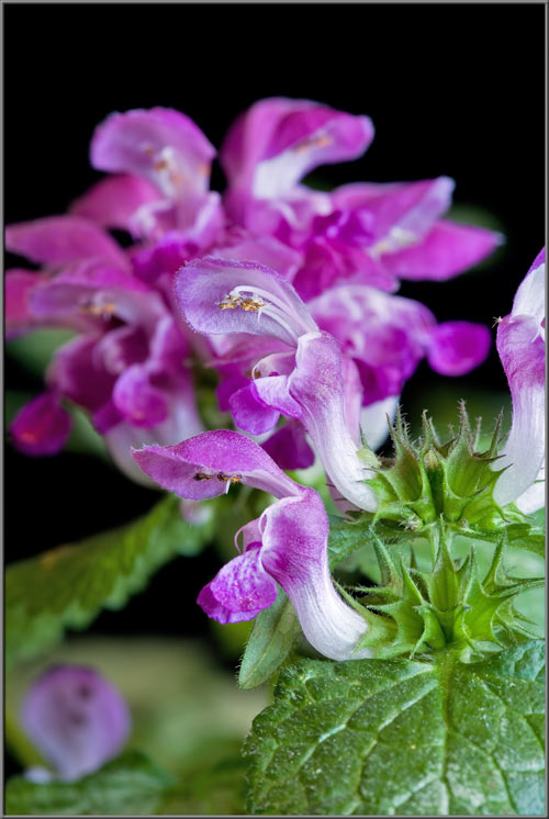
Lamium maculatum flowers grow in clusters at the tips of stems.  The flowers are remarkably similar to those of the snapdragon where the upper of the five fused petals is particularly well developed, and hangs above the rest of the flower like a hood.


A side view of the flower’s corolla reveals the two lobes that form a ‘tongue’ at the front of the flower.  Just beneath the hood are the reproductive organs, the stamens and pistil.


The bright white base of the corolla tube curves elegantly into the green cup formed by the sepals.


At the distance viewed in the two images below, it is just possible to resolve the four stamens and single forked pistil beneath the hood.  Later we will see much clearer views of these structures.


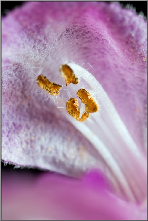
As mentioned earlier, all of the bud, and petal surfaces are particularly hairy (hirsute).


Photomicrographs showing these hairs can be seen below.  The third image shows the tiny bumps that cover their surfaces.


Only if a flower is observed from below are its reproductive structures visible.


Each flower possesses four dark anthers covered with yellow pollen.  The filaments supporting these anthers are joined to the top of the fused section of the corolla tube about at the point where the colour changes from red to white.


A closer view of an anther shows the fringe of colourless hairs that ring its dark body.


Side views show the point of connection of the filament to the anther.


A slight change in focus gives a partial view of the flower’s stigma (white – pointing down in the second image).


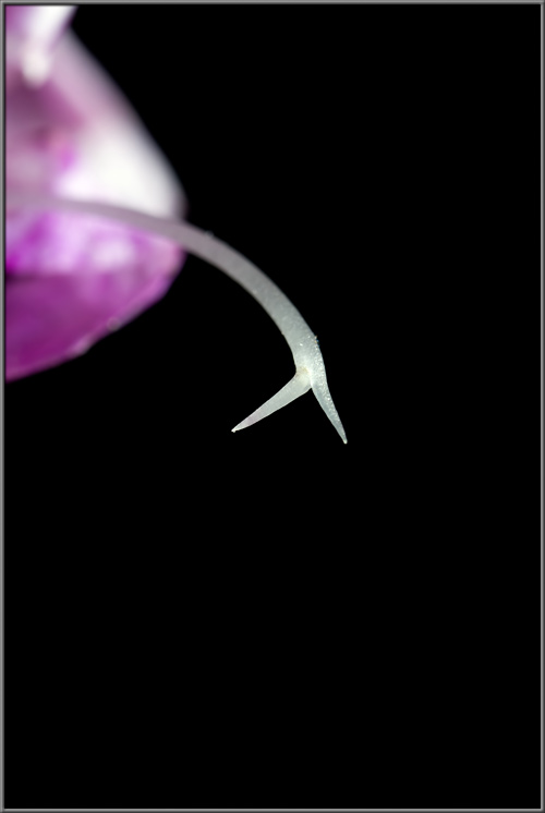
At the point where the hood meets the top of the corolla tube, there are two ‘shoulders’.  Notice the white, hanging, thread-like structure that is positioned at the point of each shoulder.  These structures also occur on several species of orchids.


The images that follow show the four anthers in a flower from different points of view.


Photomicrographs follow that show side views of an anther.


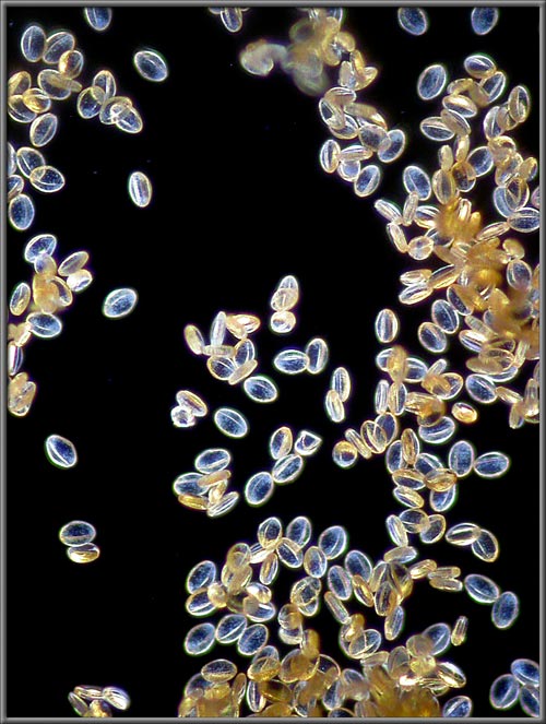
Higher magnifications allow clearer views of ellipsoidal Lamium maculatum pollen grains on the surface of an anther.


Photomicrographs showing a dusting of grains on a microscope slide reveal surface details including the longitudinal groove that bisects each grain.


The upper portion of the colourless filament, which supports an anther, is also pollen covered.


In the image on the left, one colourless, pointed lobe of the flower’s stigma is just visible.  Other blooms, like the one shown on the right, have their reproductive structures positioned deeper within the hood.


Since the pistil has a colour similar to nearby structures, it is often difficult to distinguish the forked stigma against the background.


Removing the hood makes it (somewhat) easier to see the flower’s pistil.  Notice that the stigma’s  position is roughly level with the anthers, which increases the possibility of self-pollination.  The base of its long supporting style is connected to the ovary positioned at the very bottom of the corolla tube.


Stigmas in most flowers have tiny hair-like projections that help to capture pollen grains.  This seems not to be the case in this species based on the photomicrograph at right, below.


Plant stems are sturdy, and possess relatively deep grooves along their length.  Fine hairs grow profusely on their surfaces.


Lamium maculatum’s shallowly lobed, heart-shaped leaves are one of the plant’s most admired details.  Silvery-green, with a darker green edge, they have a complex and interesting vein pattern.  Even the random, reddish-brown areas are attractive, although I believe that they may be signs of a problem.


Closer views of the upper surface of one of the leaves can be seen below.


The cellular structure of a leaf’s upper surface is visible in the two photomicrographs that follow.


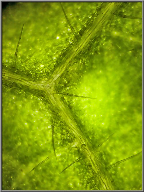
Below are photomicrographs showing the hairs growing from the lower surface of a leaf.


This strange looking sucking insect was feasting on the leaf while I was working with it.  Its size was so small that even looking carefully with the naked eye, it was barely discernable.


This deadnettle cultivar is used as groundcover in partly to fully shaded areas.  Although its flowers are not large, the overall impression given by the plant is spectacular.


Photographic Equipment


The low magnification, (to 1:1), macro-photographs were taken using a 13 megapixel Canon 5D full frame DSLR, using a Canon EF 180 mm 1:3.5 L Macro lens.

A 10 megapixel Canon 40D DSLR, equipped with a specialized high magnification (1x to 5x) Canon macro lens, the MP-E 65 mm 1:2.8, was used to take the remainder of the images.

The photomicrographs were taken using a Leitz SM-Pol microscope (using a dark ground condenser), and the Coolpix 4500.


A Flower Garden of Macroscopic Delights

A complete graphical index of all of my flower articles can be found here.


The Colourful World of Chemical Crystals

A complete graphical index of all of my crystal articles can be found here.


 All comments to the author Brian Johnston are welcomed.

Microscopy UK Front Page
Micscape Magazine
Article Library


© Microscopy UK or their contributors.

Published in the March 2013 edition of Micscape.
Please report any Web problems or offer general comments to the Micscape Editor.
Micscape is the on-line monthly magazine of the Microscopy UK web
site at Microscopy-UK  


© Onview.net Ltd, Microscopy-UK, and all contributors 1995 onwards. All rights reserved. Main site is at www.microscopy-uk.org.uk with full mirror at www.microscopy-uk.net .