A Close-up View of the

Pink Masterwort


Astrantia major 'Buckland'


by Brian Johnston   (Canada)

Astrantia major, (Greater Masterwort, or “Hattie’s Pincushion” as it was called in the past), is a member of the Apiaceae family.  Since family members have flower clusters in which each flower is joined by its stalk to a common point, they are commonly referred to as Umbelliferae.  Carrots, parsley, and celery are edible vegetable members of the family, and so is the extremely poisonous spotted water-hemlock – the most toxic plant in North America.

Amazingly, Astrantia major has been cultivated since the 1500’s.  This is probably due to the fact that it is very hardy, and produces seeds more freely than other species.  The plant’s leaves first appear in March, and its blooms appear late in May.  The name Pink Masterwort is used to describe the particular hybrid photographed for this article.

The two images that follow show what appears to be a group of flowers connected by their stalks to the top of the main stem.  Appearances can be deceiving!  In fact, each of the ‘flowers’ is not a flower at all, but instead a flower-head composed of very many tiny flowers.  What appear to be the flower’s petals are actually a whorl of bracts surrounding the cluster of real flowers.


During the late bud-stage of a flower-head, the cluster of tiny buds is partly enclosed by these petal-like bracts.  Two of these early-stage flower-heads can be seen near the bottom of the image below.


The side view that follows reveals the dramatic changes in appearance of the flower-head as it blooms.  It is evident that the “Pink” in the common name Pink Masterwort refers to the colour of the stalks supporting each of the individual flowers in the cluster.


A very distinctive feature of the species is shown below.  Although the inner surface of each bract is completely white, the outer surface displays a single, radial, green vein with short branches along its length.  Viewed from above, as in the second image, the many unopened flower buds can be seen clearly.


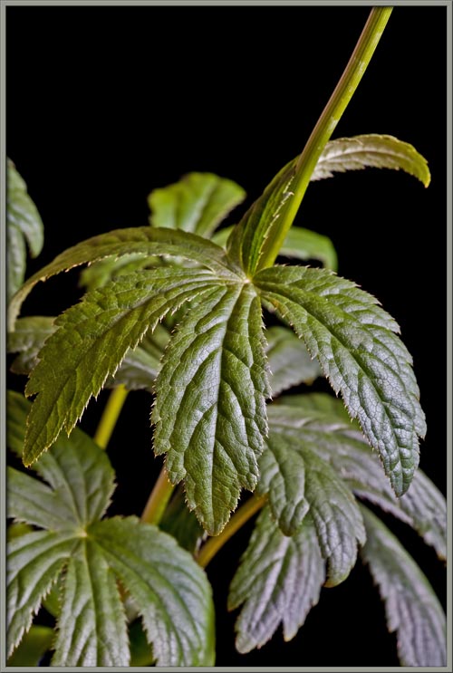
The plant’s strong stem supports many attractive green leaves, each of which possesses five palmately arranged lobes with serrated edges.


Glossy green in appearance, the upper surface of a leaf has an intricate vein pattern, and an edge with tiny, needle-like projections as part of the serration.


The under-surface of a leaf is much rougher, and is much lighter in hue.  In fact, under the lighting conditions of the photograph, it appears almost white, thus increasing the contrast with the darker green vein structure.


Notice, in the image below, that the lower of the two flower-heads is at a later developmental stage.  Not only is it larger, but out of each tiny flower there project two hair-like stigmas.


At the point where the stalks of the flower-heads join the main stem, there is a whorl of small, greenish-white leaflets.


These four leaflets can be seen in the two images that follow.


Unlike the bracts surrounding the flower-head, the leaflets have three parallel green veins, each with many smaller off-shoots.  The colour scheme of leaflets and bracts is identical.


Even the stalk supporting the flower-head is unusual.  Its shape is not cylindrical, but rather cusp-like, with the channels a green colour, and the longitudinal ridges a white colour.


Under the microscope, the cells making up the surface and veining of a leaflet become visible.


Higher magnification photomicrographs showing the tip of a leaflet (left), and the underside surface (right) can be seen below.  Notice the prominent stomata and guard cells that control gas access to the interior of the leaf’s underside in the right-hand image.


As an Astrantia major flower-head matures, its bracts flatten and open out to reveal the many flowers within.  These flowers mature from the centre of the group outwards.  This is indicated in the photograph by the emergence of the hair-like stigmas and styles mentioned earlier.


Notice in the two images that follow, the green tips of the bracts that frame the flower-head, and their dark pink bases.  The stalk of each flower is also pink.


Most of the flowers in the images below are immature, but a few have their two stigmas and styles protruding.  If you look closely, you can also see five, white, rod-like structures that curve from the periphery of the flower to its centre.  These are the stamens’ supporting filaments.  (The anthers are hidden within the body of the flower itself.)


A red anther, (male pollen producing organ), and its supporting filament can be seen more clearly in the photomicrographs below.


The cellular structure of the tip of one of the flower’s two stigmas (female pollen accepting organ), and its supporting style can be seen in the photomicrograph that follows.


If one removes some of a flower-head’s bracts, and several of its flowers, the extraordinary structure of an individual flower is revealed.  Starting from the bottom, each flower is supported by its pink stalk.  The stalk is connected to a strangely shaped, pale green, barrel-shaped ovary (seed producing organ).  At the top of the ovary are five tooth-shaped sepals (modified leaves), each of which is dark green and has a pale pink edge. Visible between these sepals are the five, paler pink petals.  Above these structures are the filaments, and in some cases, the two pistils.  (You may also notice that wherever the filaments have been severed, a white, sticky liquid is exuded.)


An ovary like the one seen below, which is positioned beneath the flower’s sepals and petals, is referred to as inferior.  The intricate structure of the ovary’s pale green longitudinal ribs will be seen more clearly later in the article.


Higher magnification macro-photographs showing a flower’s outer whorl of green sepals, and inner whorl of pale pink (almost white) petals can be seen below.


Very high magnification macro-photomicrographs of the sepals and petals of a flower reveal more details.  (The whorl of green sepals is referred to as the flower’s calyx, while the whorl of petals is referred to as the flower’s corolla.)


Below are two photomicrographs showing the cellular structure of the surface of the ovary.  The ridges take the shape of a meandering river.


Higher magnification photomicrographs follow that show the cells forming a section of the ovary’s ridges.


Notice that at the very top of the ovary, the ridges are tooth-shaped.


Astrantia is native to Balkan coppices (thickets), and sub-alpine meadows.  Over many years it has been successfully trans-located to grow in gardens almost everywhere.  Its charms sadly, are not seen at normal viewing distances.  When viewed close-up however, its spectacular, intricate structure is fully revealed!


Photographic Equipment

The low magnification, (to 1:1), macro-photographs were taken using a 13 megapixel Canon 5D full frame DSLR, using a Canon EF 180 mm 1:3.5 L Macro lens.

An 8 megapixel Canon 20D DSLR, equipped with a specialized high magnification (1x to 5x) Canon macro lens, the MP-E 65 mm 1:2.8, was used to take the remainder of the images.

The photomicrographs were taken using a Leitz SM-Pol microscope (with a dark ground condenser), and the Coolpix 4500.


A Flower Garden of Macroscopic Delights

A complete graphical index of all of my flower articles can be found here.


The Colourful World of Chemical Crystals

A complete graphical index of all of my crystal articles can be found here.


 All comments to the author Brian Johnston are welcomed.


Microscopy UK Front Page
Micscape Magazine
Article Library


© Microscopy UK or their contributors.

Published in the March 2009 edition of Micscape.
Please report any Web problems or offer general comments to the Micscape Editor.
Micscape is the on-line monthly magazine of the Microscopy UK web
site at Microscopy-UK  


© Onview.net Ltd, Microscopy-UK, and all contributors 1995 onwards. All rights reserved. Main site is at www.microscopy-uk.org.uk with full mirror at www.microscopy-uk.net .