SUCTORIA
by Jean-Marie Cavanihac, France
Note: This article contains some animated images which may take some time to load. Two of them are shown via a link to a separate page. 
The microscopic world often contains strange forms of life. Sometimes, I think that the horror movie makers should look closer in our microscopic zoo as a source for their ideas and to imagine incredible or frightening extraterrestrial creatures. They just have to multiply their size by some thousands of times!
Some of these creatures are really tiny vampires. They are suctorians, sessile protists, Phylum Ciliophora, Class Kinetofragminophora; some of them live in a rigid house called a lorica, and the main species affix to objects like algae by a non-contractile stalk. These protozoa do not have a cytostome (mouth) to eat their prey but do possess two bundles of straight tentacles - each one terminated by a rounded structure like a disk and probably adhesive. It's astonishing to see how these tiny structures are slowly deployed from the rigid body. An animated picture is shown below.


Click HERE to see an animated picture.

Some pictures of suctorians are shown below:
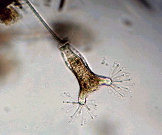
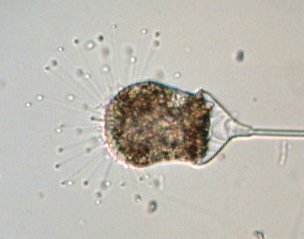
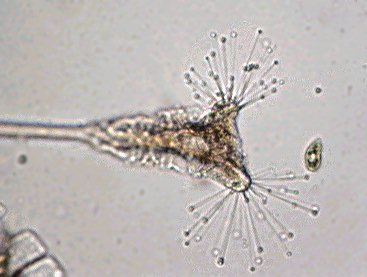
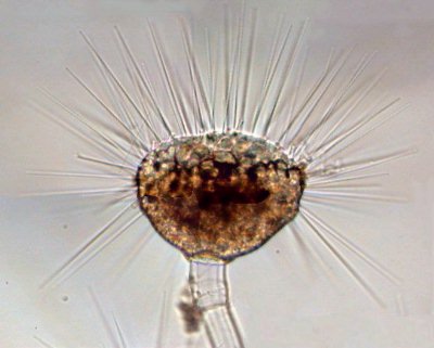
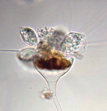
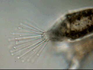
When another protozoan touches one of these sticky disks it remains glued and immediately other tentacles are brought into play to immobilize it. Then a strange ballet begins if you observe carefully: first, disks seem to dissolve the protozoan cell membrane, then a sort of granulated flow is seen along each tentacle from protozoan toward the suctoria, as if the protozoan cytoplasm was transferred little drops by little drops. In a few minutes, the entire cytoplasm of the prey is phagocytized through the suctorian tentacles! At the same time, cilia of the protozoan stop moving, and some minutes later, an empty and retracted membrane of the protozoan is rejected. You don't believe that is possible? Look at the animated picture shown right taken in only five minutes of real time!
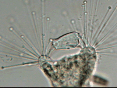
 
 
All the specimens shown here are marine organisms. I found them fixed on one species of bryozoan, red colored, and only on those that were near the water surface. I never encountered them on white bryozoans having a chalky structure.
Go to page 2 : To see a strange metamorphosis
Comments to the author Jean-Marie Cavanihac are welcomed.

Microscopy UK Front Page
Micscape Magazine
Article Library


All drawings and photographs © Jean-Marie Cavanihac 2002

Published in the March 2003 edition of Micscape Magazine.

Please report any Web problems or offer general comments to the Micscape Editor,
via the contact on current Micscape Index. 

Micscape is the on-line monthly magazine of the Microscopy UK web
site at Microscopy-UK

WIDTH=1


© Onview.net Ltd, Microscopy-UK, and all contributors 1995 onwards. All rights reserved. Main site is at www.microscopy-uk.org.uk with full mirror at www.microscopy-uk.net.