|
|
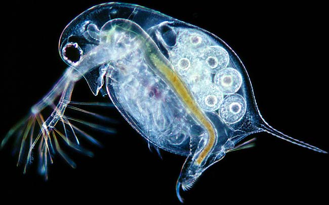
This is a mouse-over image of Daphnia longispina, a common water-flea. Use the underlying image to examine its main anatomical features! |
|
|
|
of Daphnia |
head and eyes |
ovary and embryos |
heart |
Daphnia |
| Water-fleas
are easy to find. Most ponds will provide enough of these small crustaceans.
They are ideal subjects for study under the microscope. You can examine
them swimming under a low power stereo-microscope. When observed with stronger
magnification it is best to use a deep slide. You can add dots of vaseline
to the corners of the coverslip to prevent the water-flea being damaged.
It is wonderful to see the heart beating and the blood flowing!
The body of a water flea is protected by a shell-like carapace. This looks like two valves but consists of one piece. It covers the five pairs of leaf-like legs. These are used to filter food but also bear gills. At the end of the body (abdomen) we find the post abdomen with a paired claw (furca). It is used to remove unwanted particles between the carapace. |
Find about more about them in the Micscape article Water fleas
All comments to the author Wim van Egmond are welcomed.
Visit Wims home page for links to his many web pages on microscopy
Please report any Web problems or
offer general comments to the Micscape
Editor,
via the contact on current Micscape
Index.
Micscape is the on-line monthly magazine
of the Microscopy UK web
site at Microscopy-UK