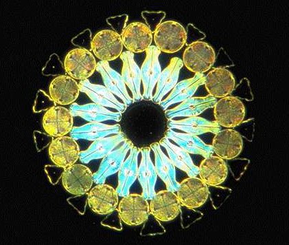
The photomicrograph below is of an arranged slide made by the skilled Klaus Kemp. Klaus is still making slides, very much so in fact. His details can be obtained from the 'Sales and Wants' section of the site. Recently he has been following in Moeller's footsteps in making some of the arranged designs that made him so famous. (See earlier article on his type slide).
Whilst the slide looks super in normal bright-field I think that it really looks best in dark-field, though photographs, especially via the computer screen can never do justice to the marvellous jewel-like effect that you get in viewing down the microscope.
So have another look at some of your favourite slides or subjects especially freshwater life, under different illumination, it is well worth it.
Editor's note:
The Micscape article Making dark-field illumination is easy describes how to simply and cheaply experiment with dark-field.
First published in June 1998 Micscape Magazine.
Please report any Web problems
or offer general comments to the Micscape Editor,
via the contact on current Micscape Index.
Micscape is the on-line monthly
magazine of the Microscopy UK web
site at Microscopy-UK