Oh yes, I still have some images that I took recently of those old slides that I haven’t yet rambled on about. So, prepare yourself and we’ll look at some more marvels and disasters from a century or more ago. In this particular batch, we have everything from Dogfish scales to a cross section of the stem of a gourd. What more could one want? Well, how about a larval starfish, a goldfinch plume, and an ovipositer of a crane fly.
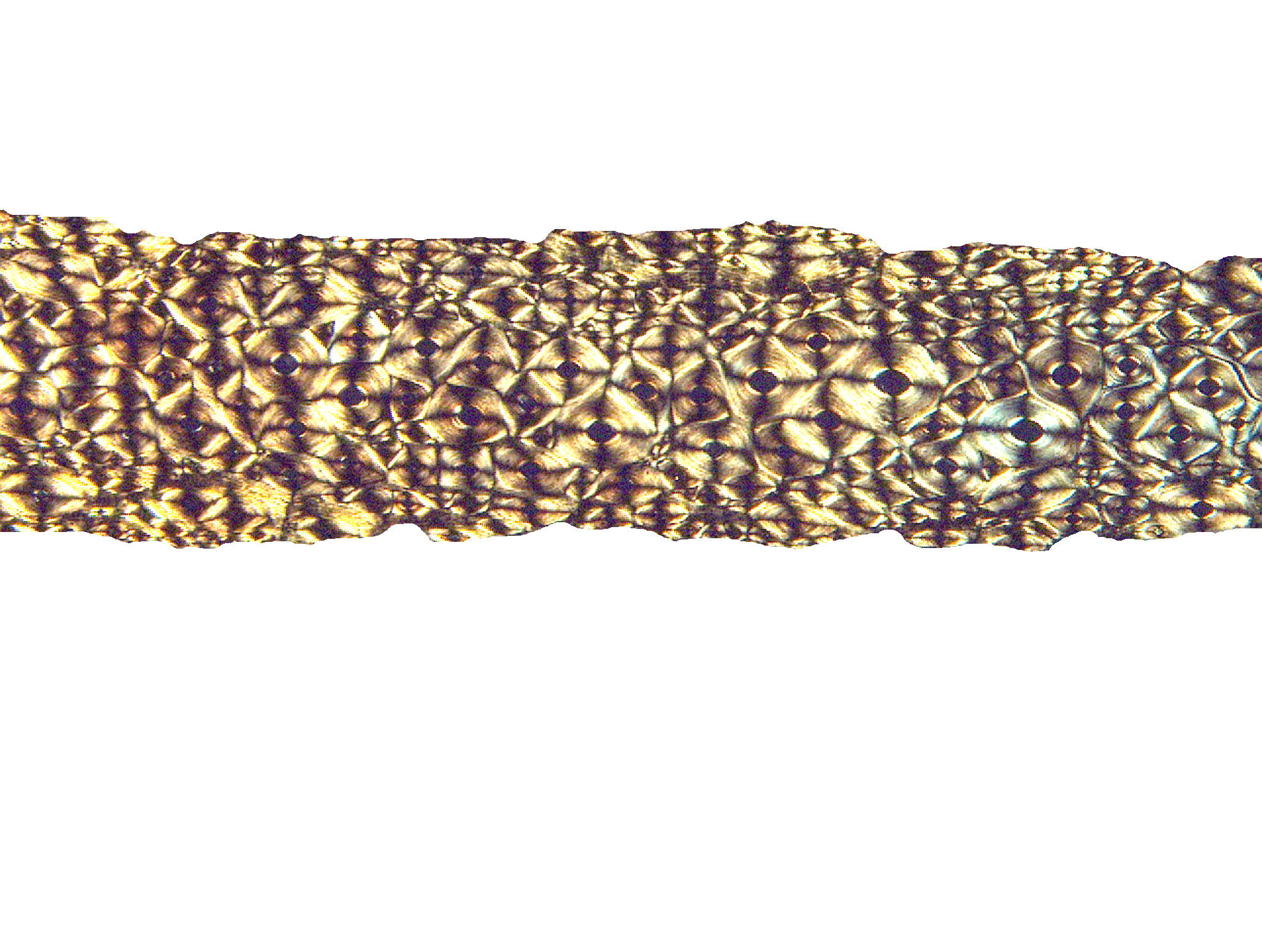
Let’s begin with the dogfish scales. The dogfish is a shark and the spines have points that are very sharp. The dogfish, unlike other sharks, has venomous spines in front of each dorsal fin. The placoid spines which we are looking at are tightly packed. If one runs ones hand down the back from head to tail, the surface feels smooth, but reversing direction, the texture is like that of a file or rasp.
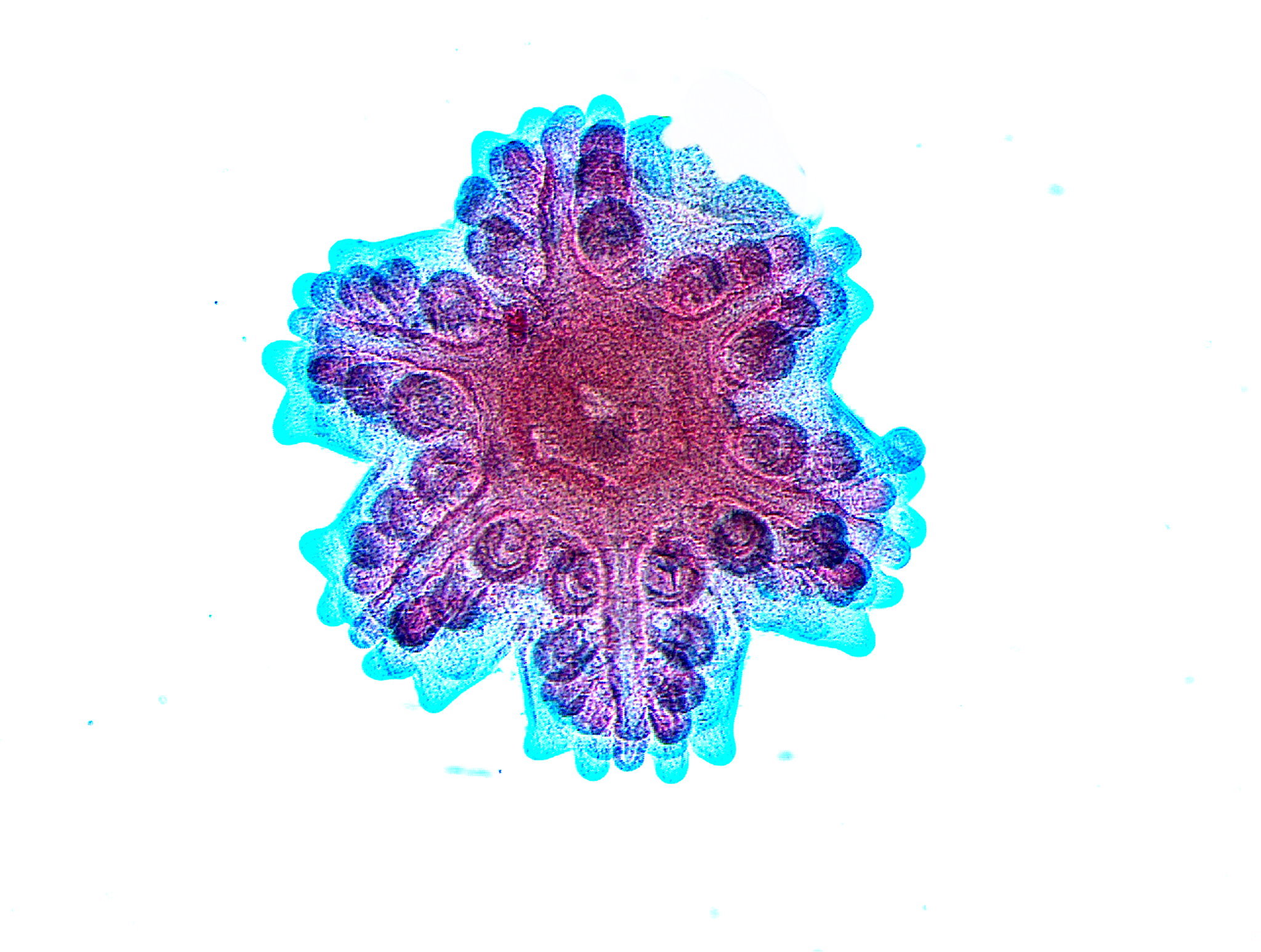
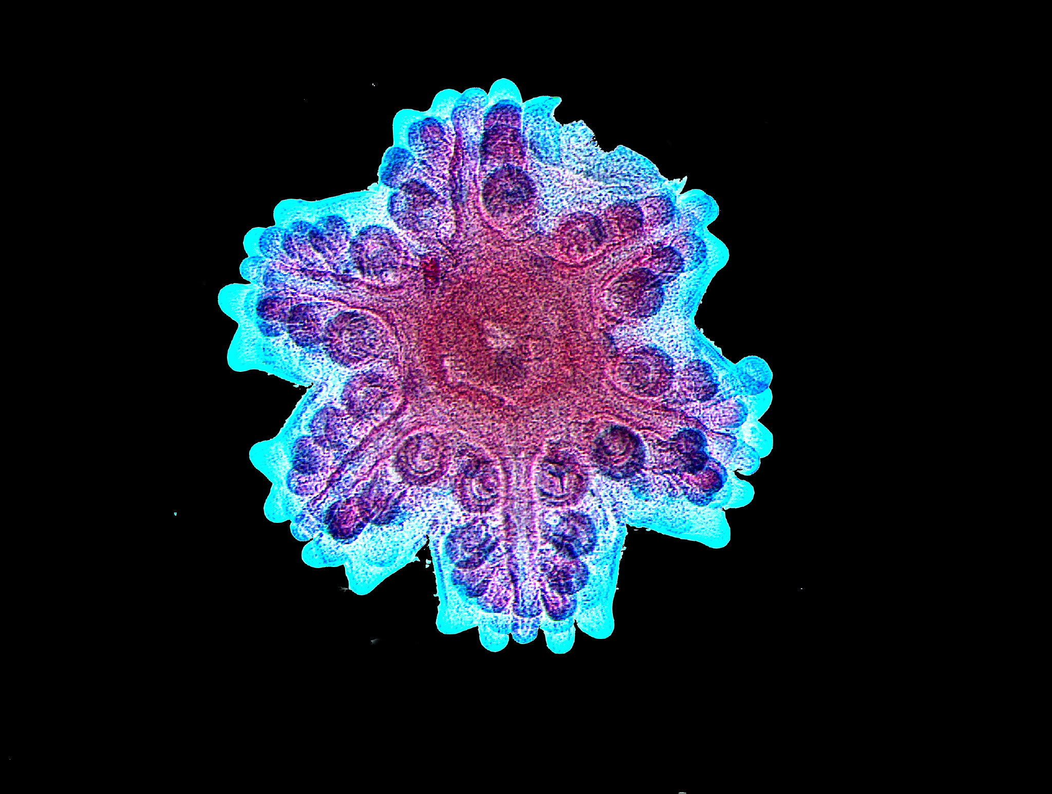
Next a very different kind of fish which isn’t a fish at all although many people call it a starfish, whereas, other more finicky types insist on saying that it’s a sea star. This is a larval stage and I’ll show you two views; the first with a white background and the second with a black background which I prefer, as it think it increases the contrast.
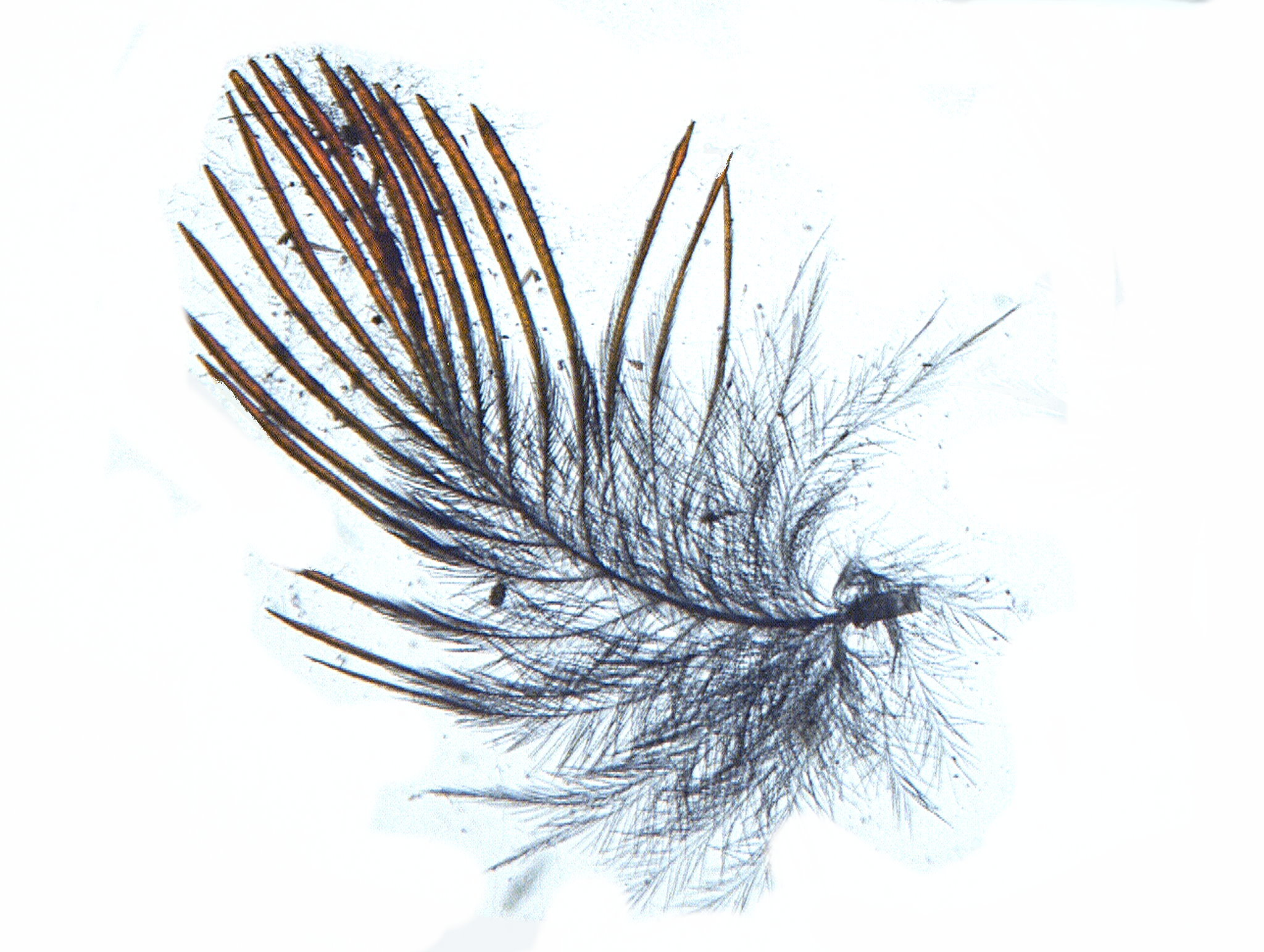
I mentioned a goldfinch plume. This was a tricky item to photograph and process. The first image is modified brightfield with a shift in background and designed to show the tint of “gold” in the plume.
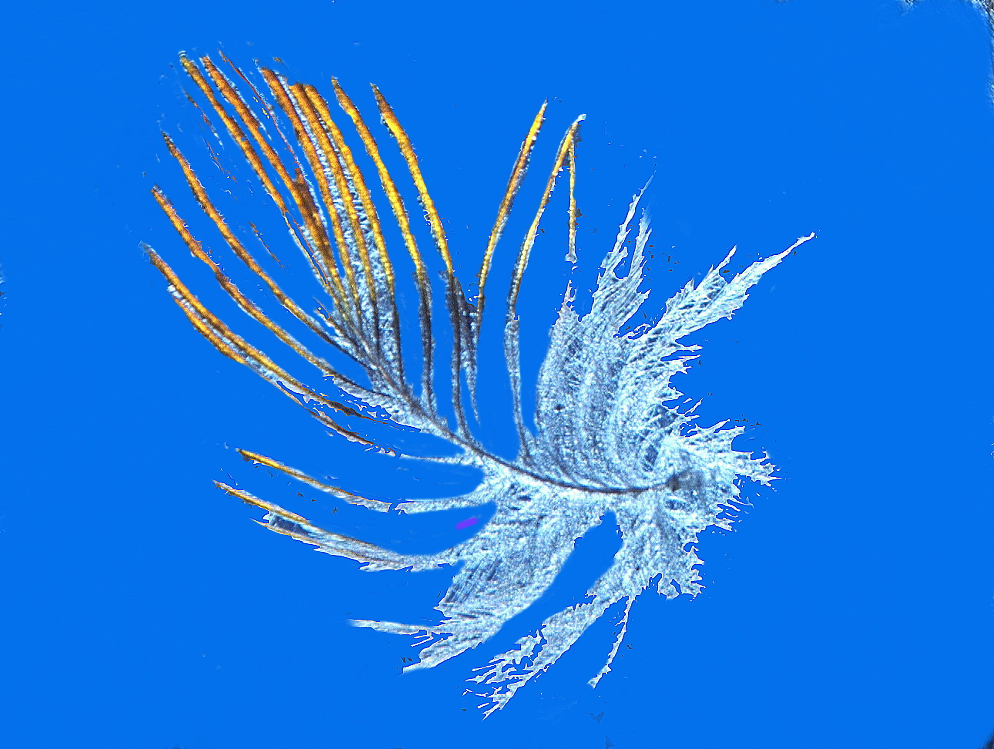
Quite lovely, isn’t it; but a better view is obtained by using oblique illumination with a white background.
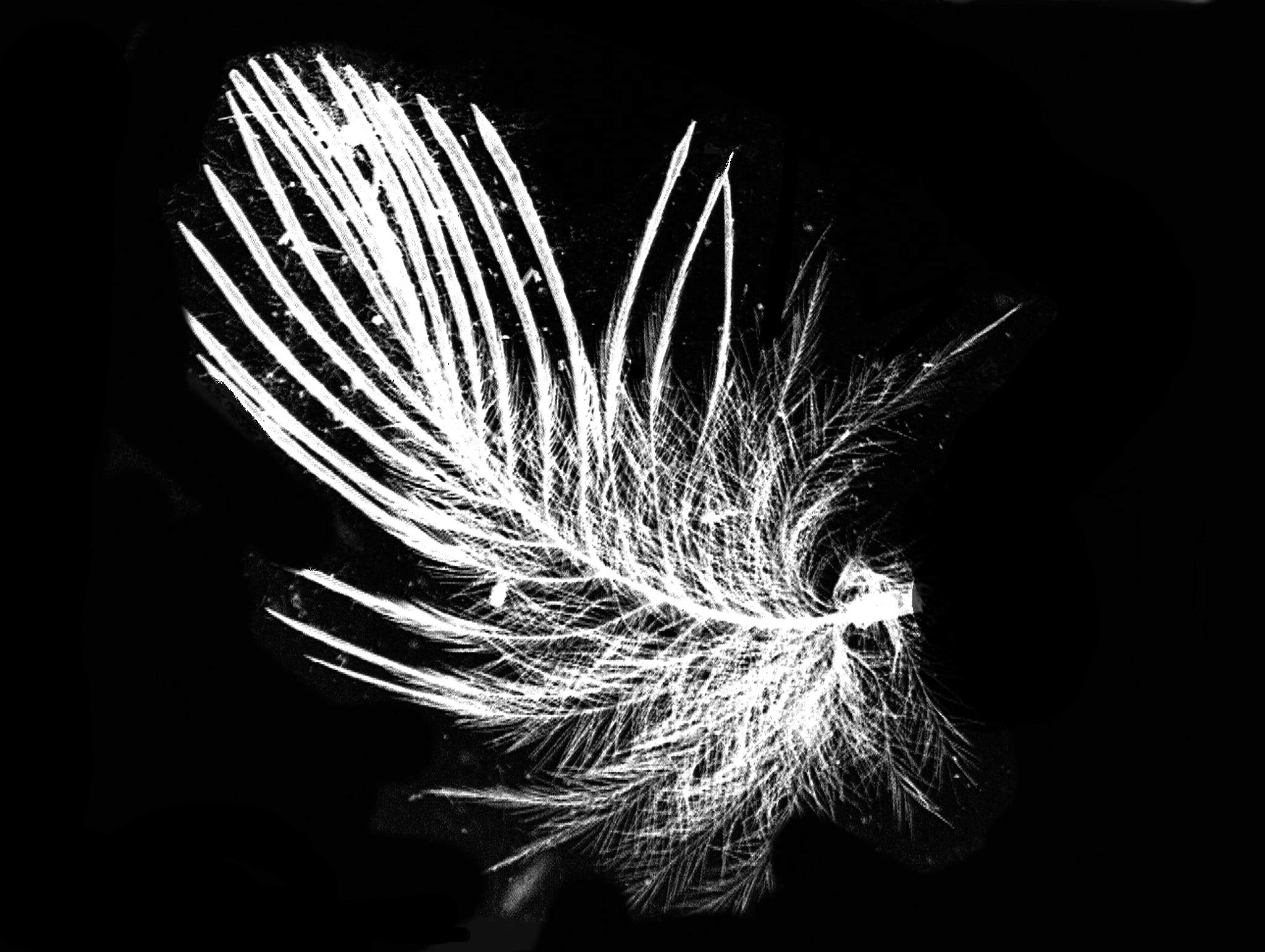
Then I took that image and applied the “invert” function and we get yet another sort of contrast.
While we’re looking at fuzzy things, here are a couple of images of the head and fuzzy antennae of a blowfly, also known as a bluebottle or greenbottle fly. The descriptor blowfly occurs in Shakespeare and is a reference to the fact that these critters would lay eggs in meat and then the larvae would feed giving it the appearance of being “flyblown” The first image is brightfield and then I applied the “invert” function to it which produced a pleasing contrast.
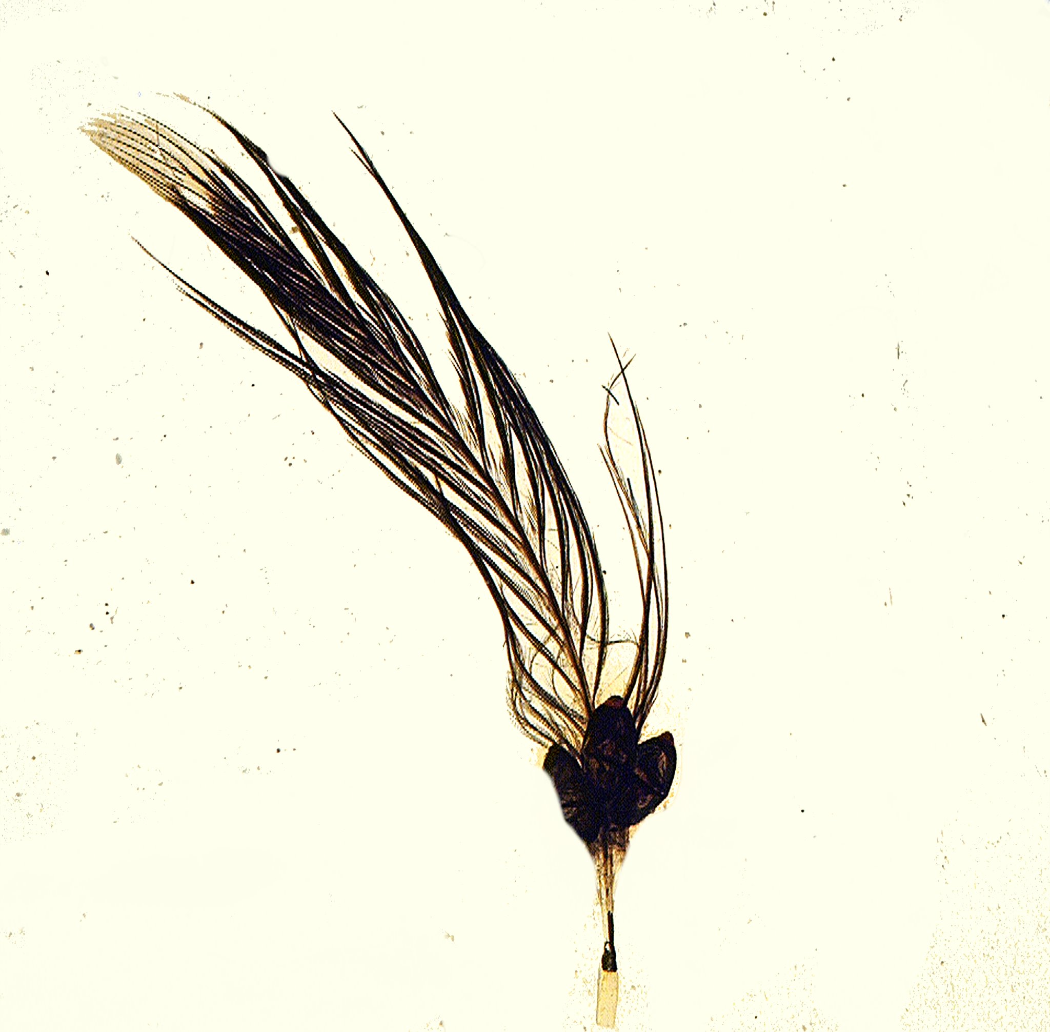
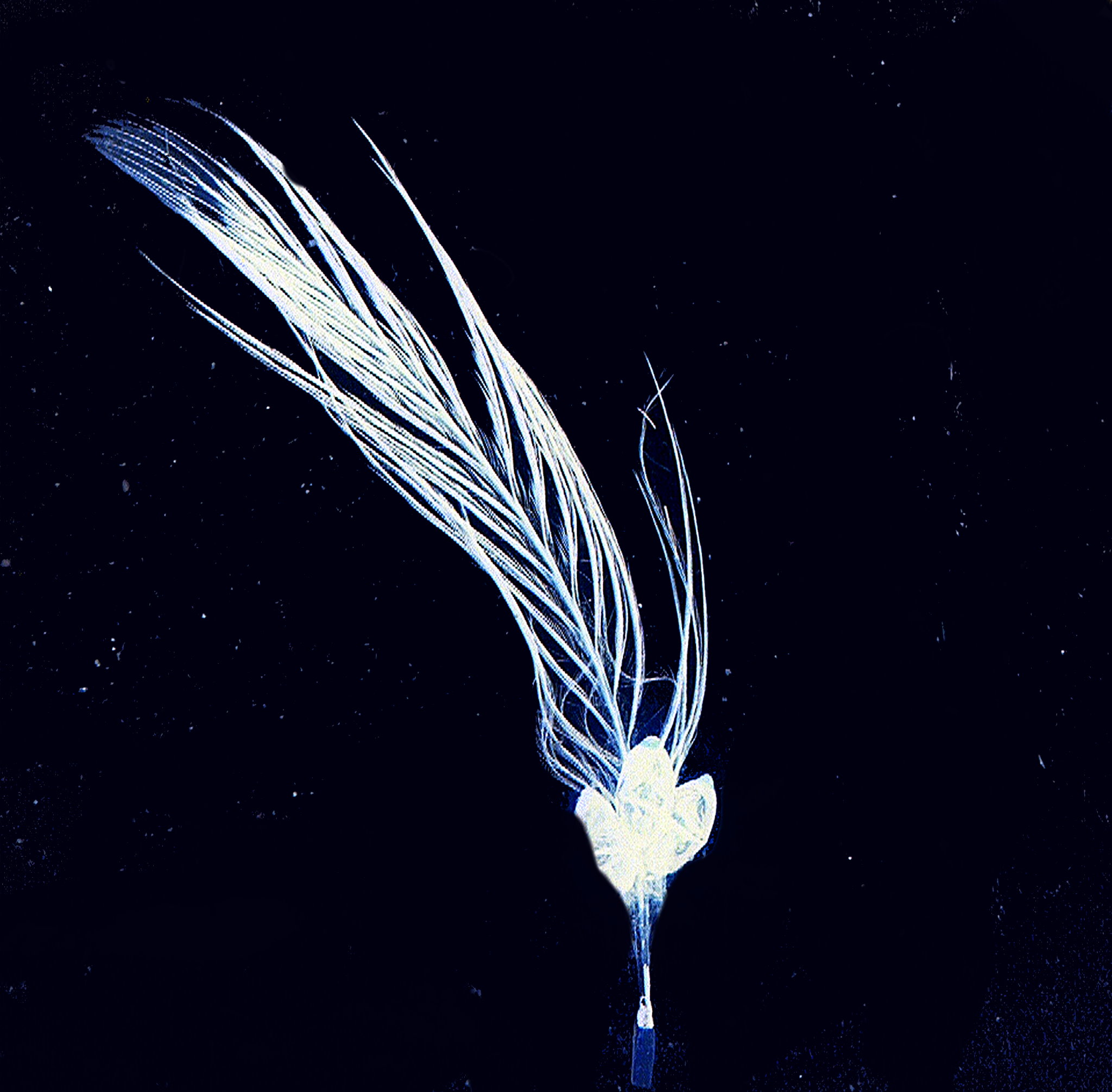
Again, I’ll show you a brightfield image and then the “invert” which quite strikingly reveals some of the internal structure. This is an example of how valuable it can be to experiment with various kinds of contrast and illumination in dealing with such images.
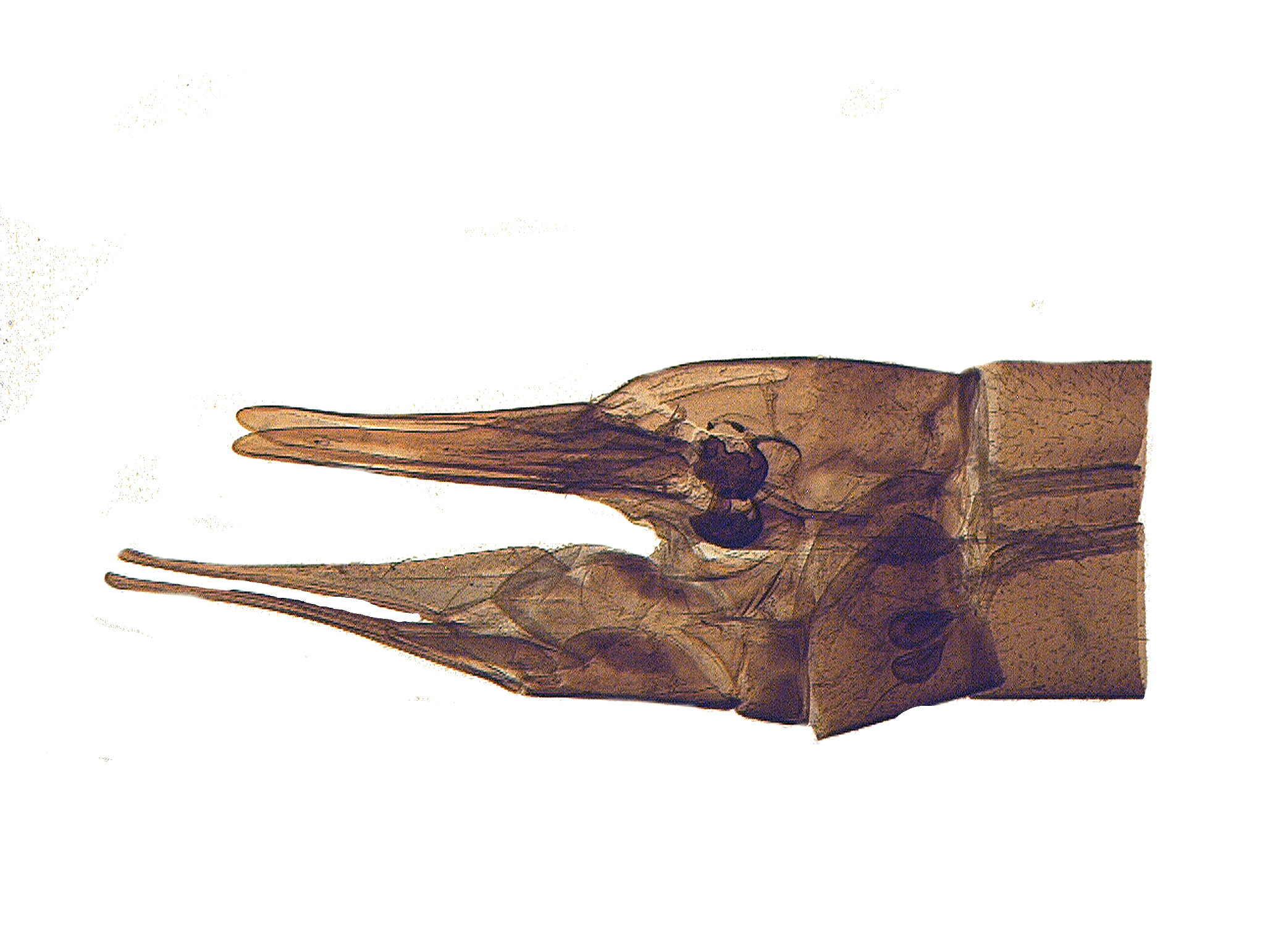
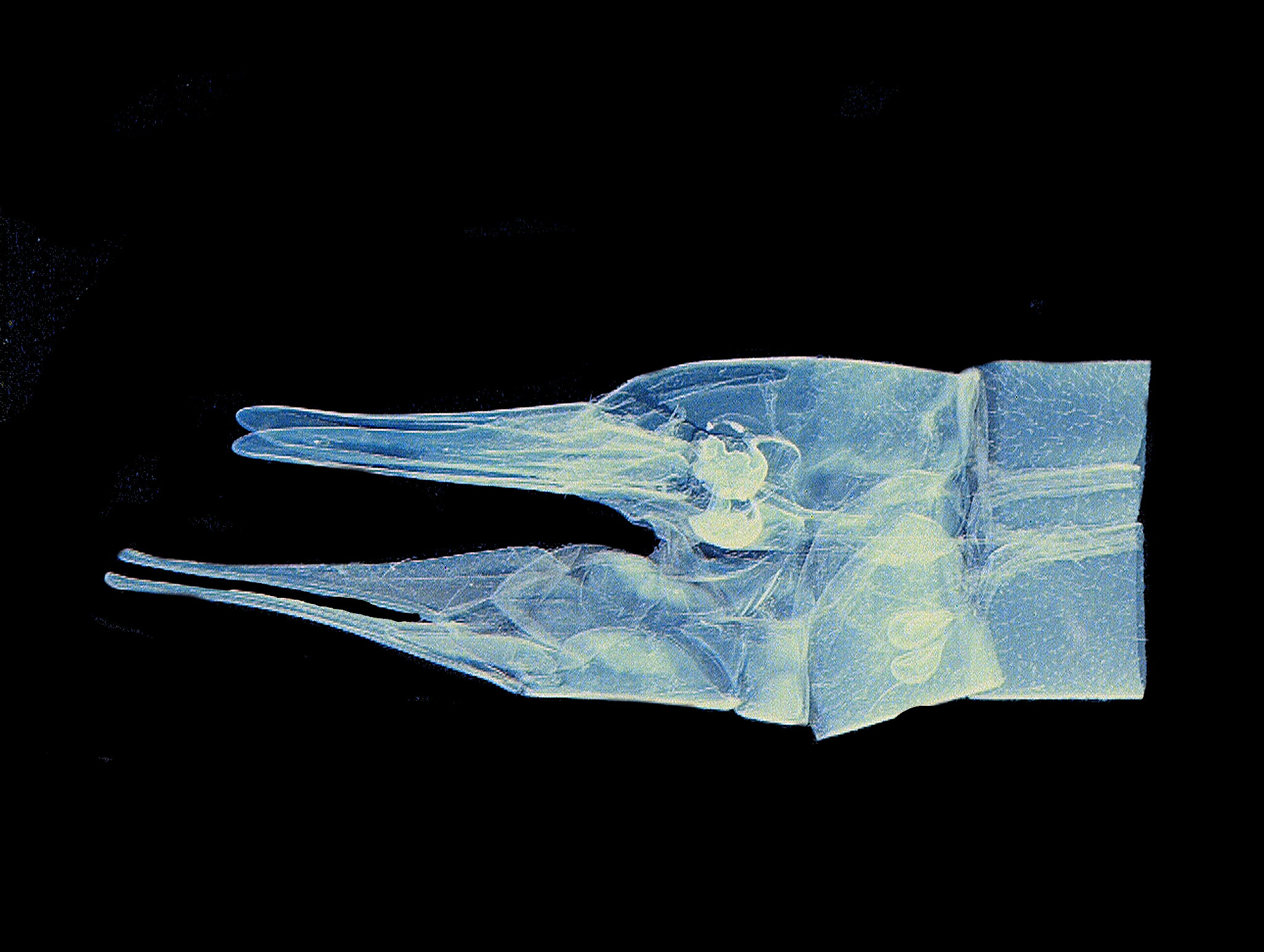
While we’re looking at insect parts, we might as well continue. These were popular subjects for 19th Century microscopists. This next image is that of the head of a beetle identified only as a “Green Beetle”. It’s hard to tell whether it’s giving a gesture of surrender or indifference.
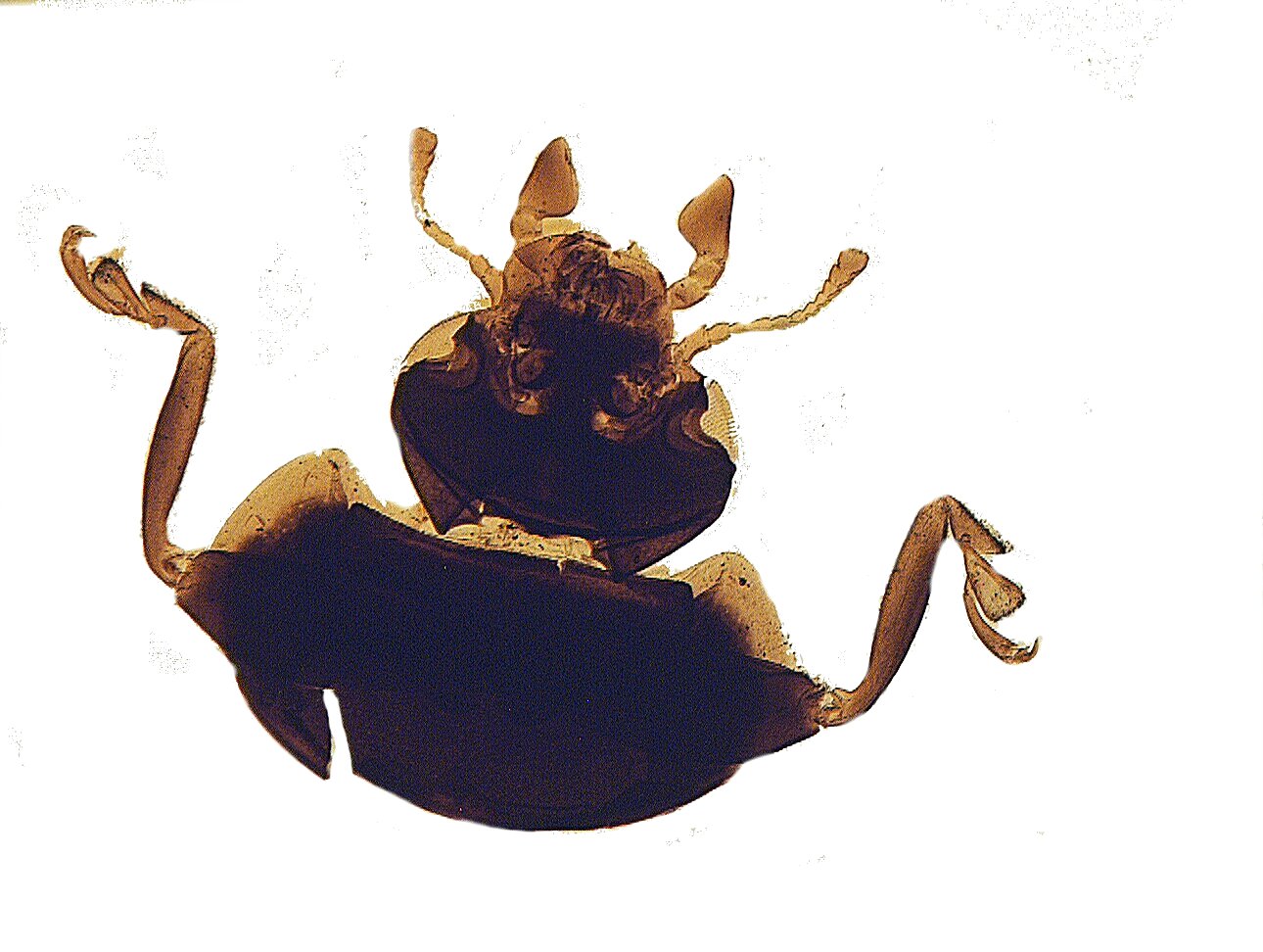
Next up is a portion of the wing of a Red Admiral butterfly. Not only can you see the various colored scales, but also an incredible number of tiny hairs along with the edges and on the surface.
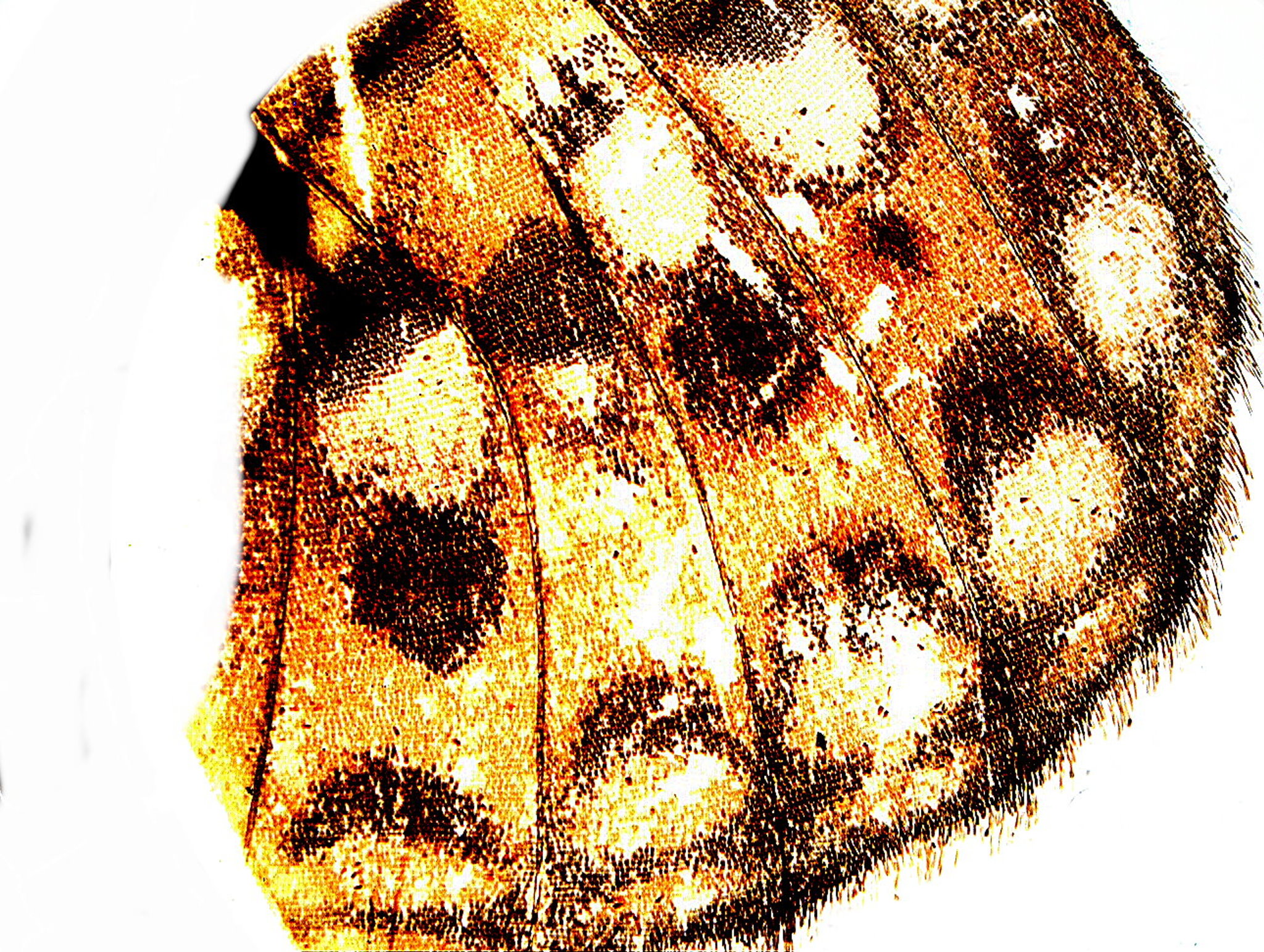
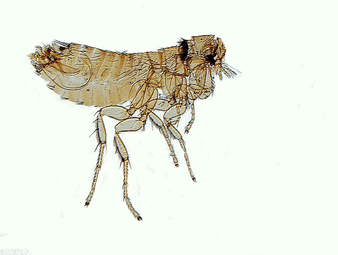
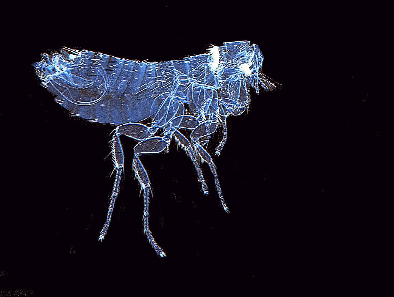
A nuisance that can go after your cats, dogs, hamsters, and even you (different species, of course) is the flea. The humorous poet Ogden Nash is reputed to have written the shortest poem in the English language which I will provide here for your delectation.
Fleas
Adam, had ‘em.
First brightfield and then invert which gives us a different glimpse by demarcating the segmentation quite distinctly.
Making a radical leap to the plant kingdom, we find a slide with what is described as a cornflower. At first glance, it almost looks like a rather pathetic, faded starfish. However, on closer examination, it is clearly botanical. The invert image (the third one) again is interesting in what it reveals of the internal structure.
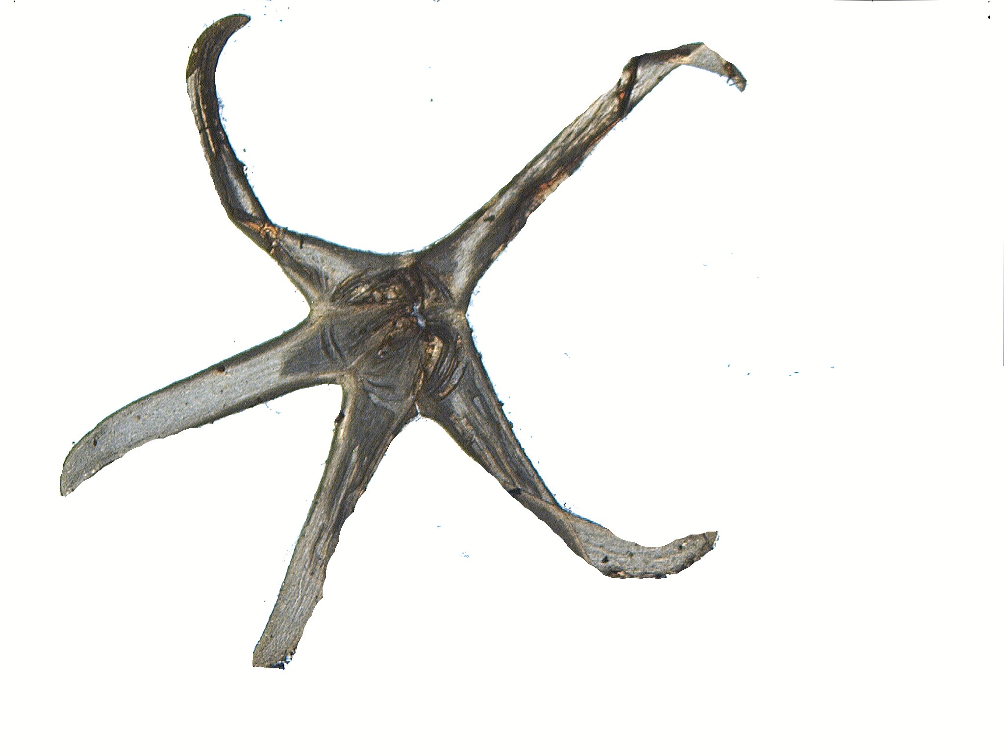
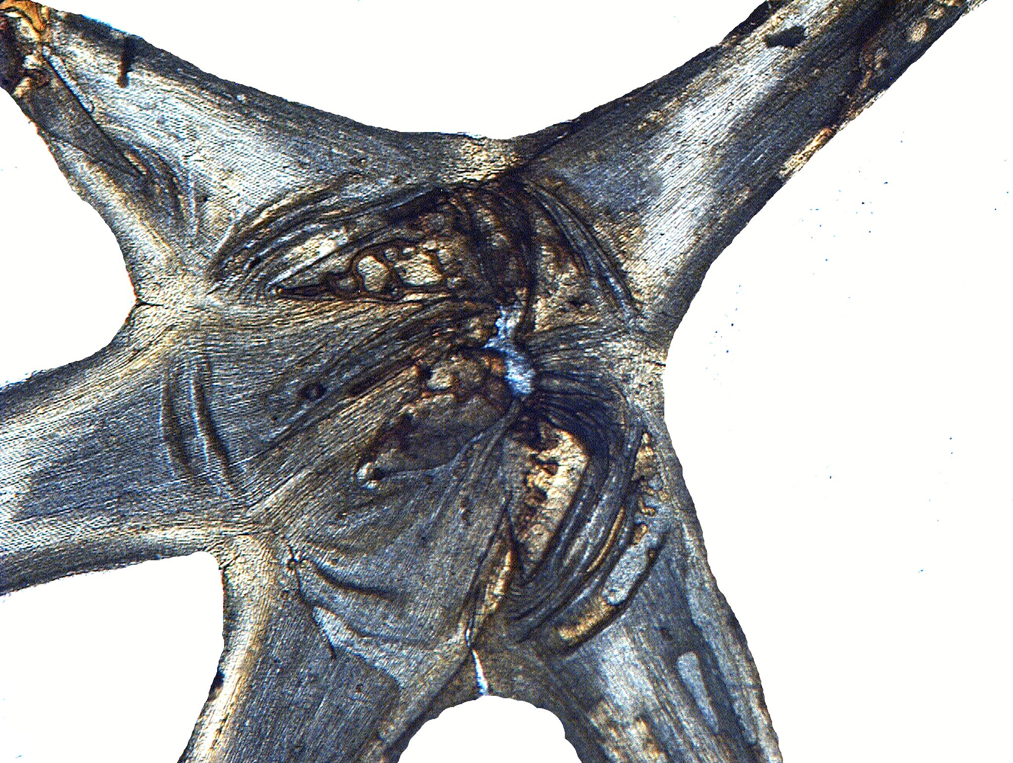
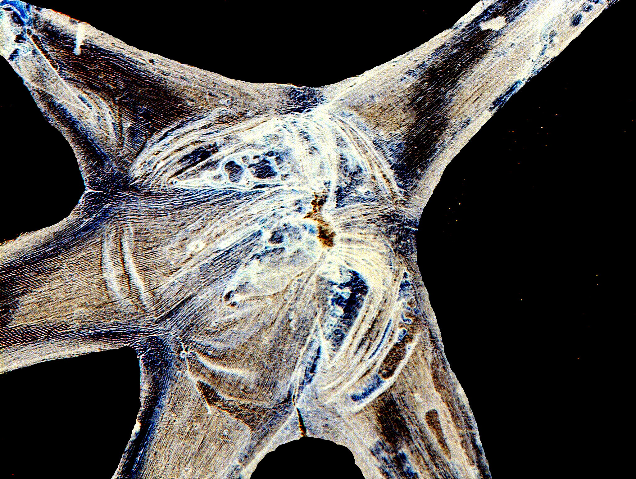
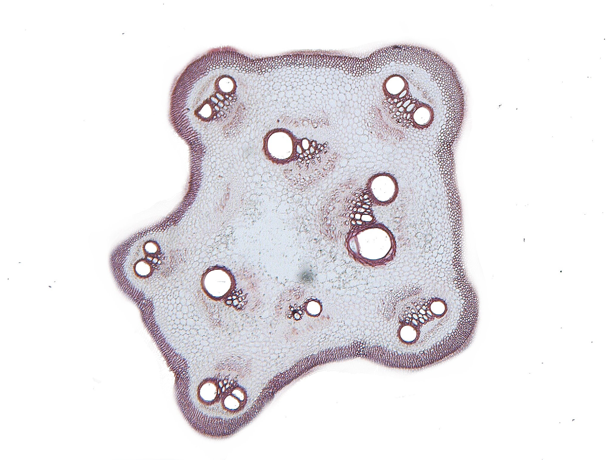
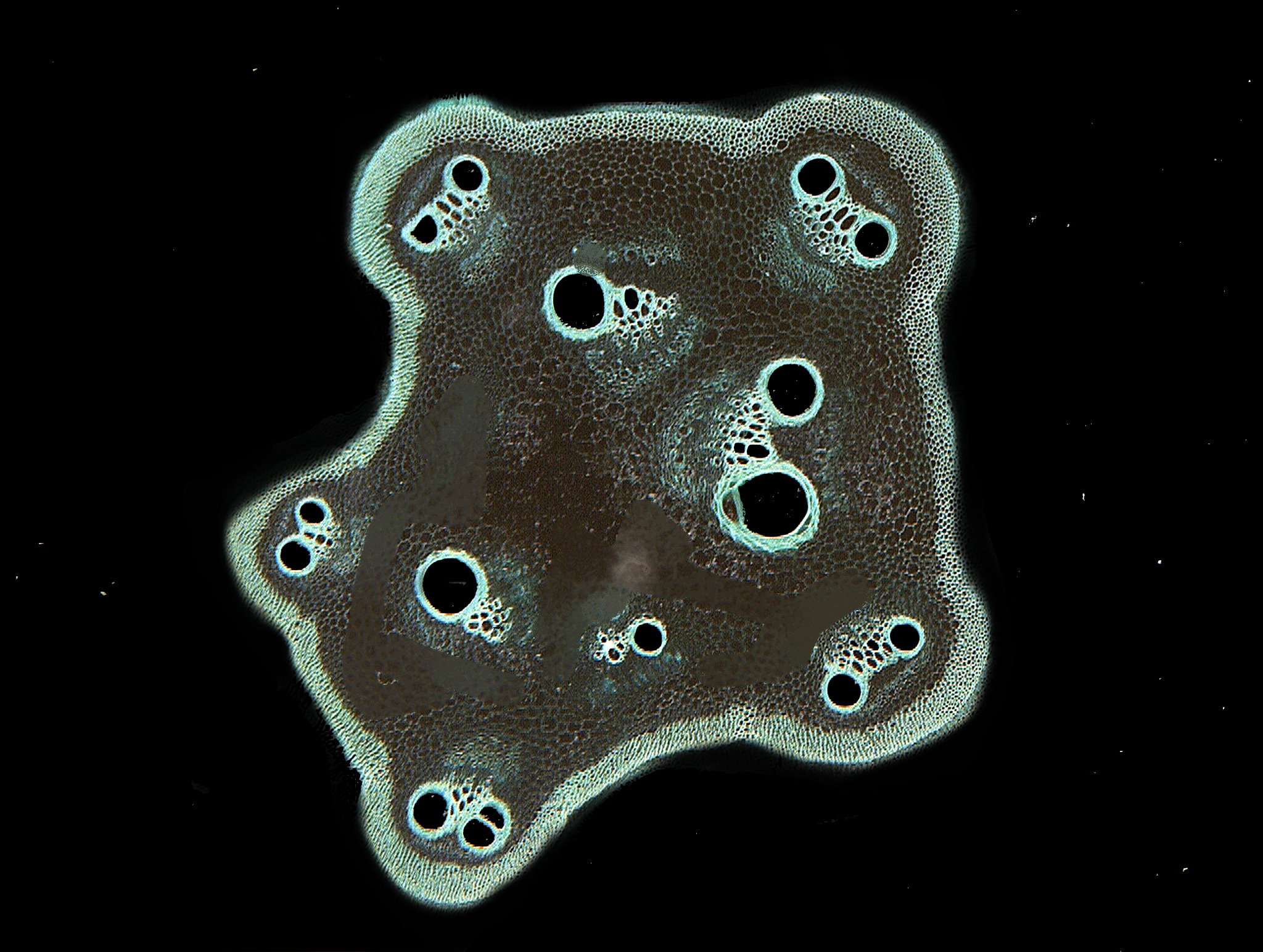
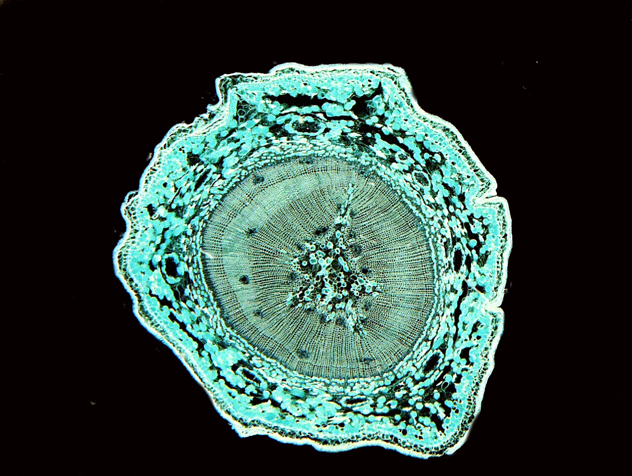
In addition, I found a nice cross section of a gourd stem. You can see both the vessels and vascular bundles. The invert image I find particularly interesting.
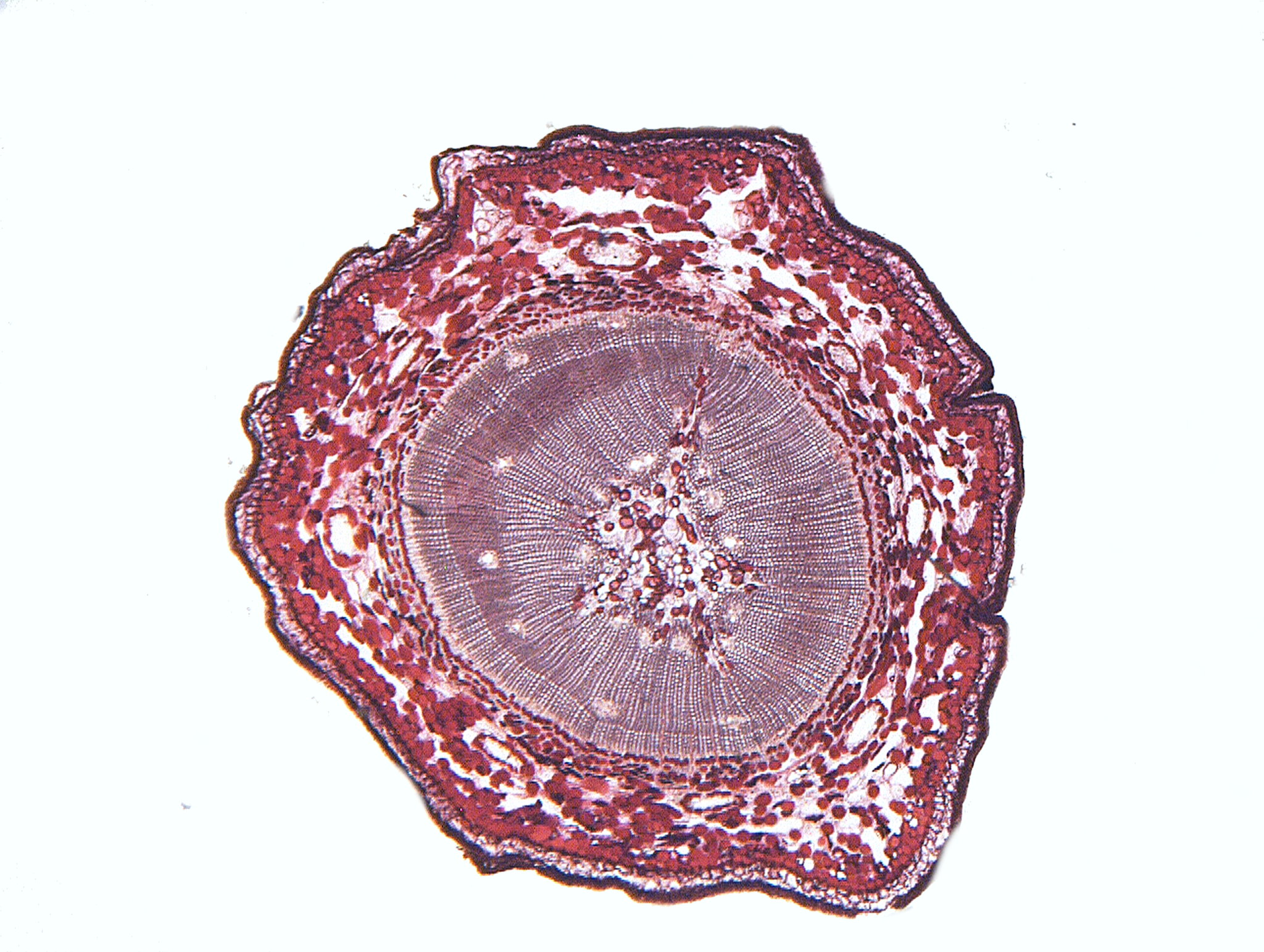
Another stem of interest is a cross section of a woody stem of pine. This is a practically new slide being only 50 or 60 years old and made in post-war Japan. Again, the invert image is interesting in the manner in which it shifts the highlights.
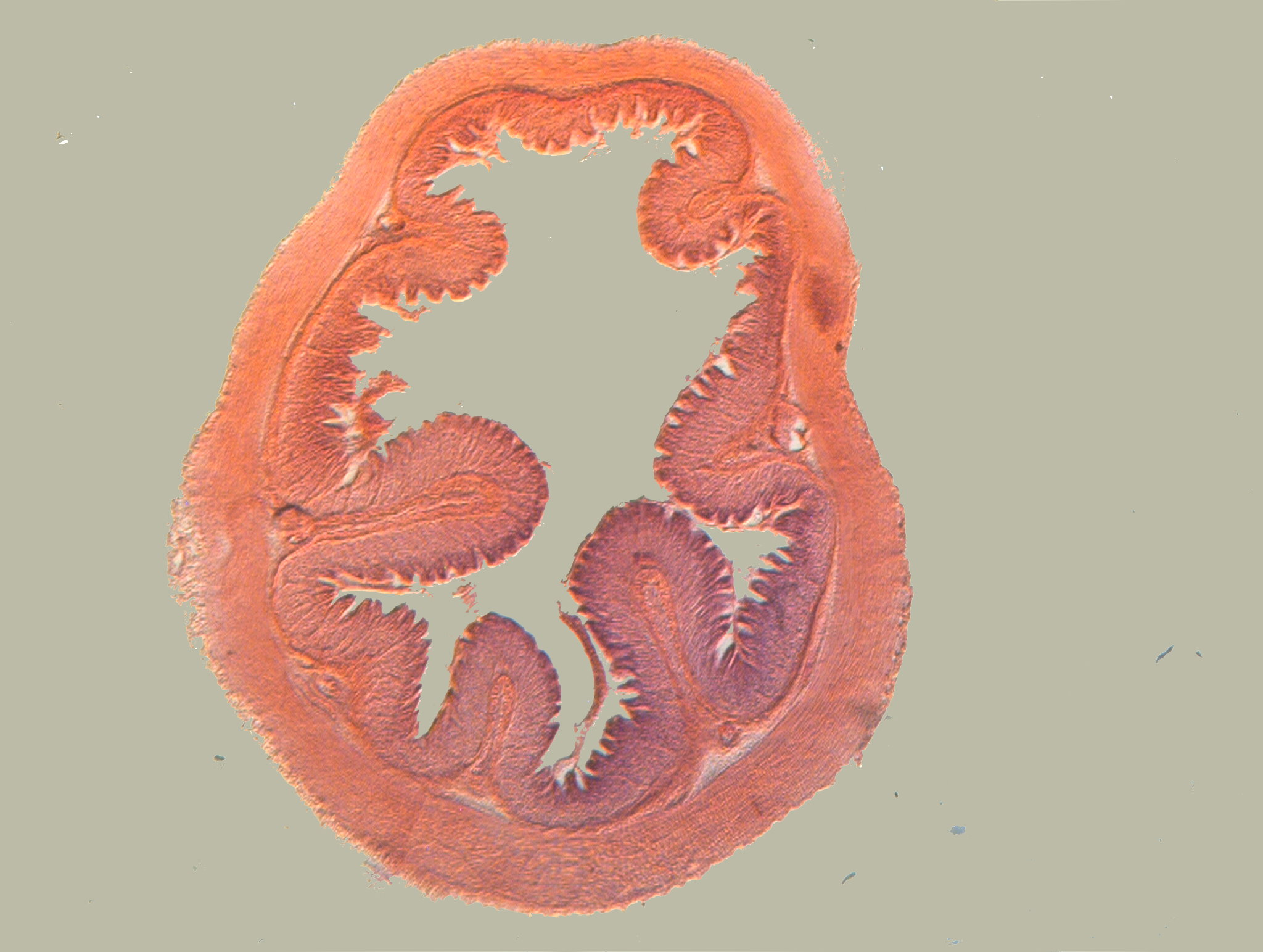
Now, for something completely different; the cross section of the stomach of a frog and remember it’s only the legs that you eat.
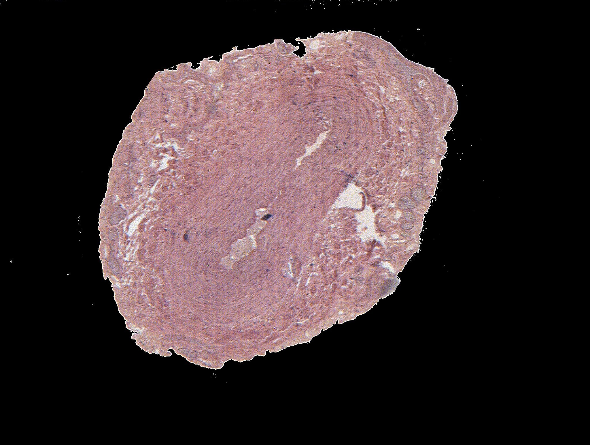
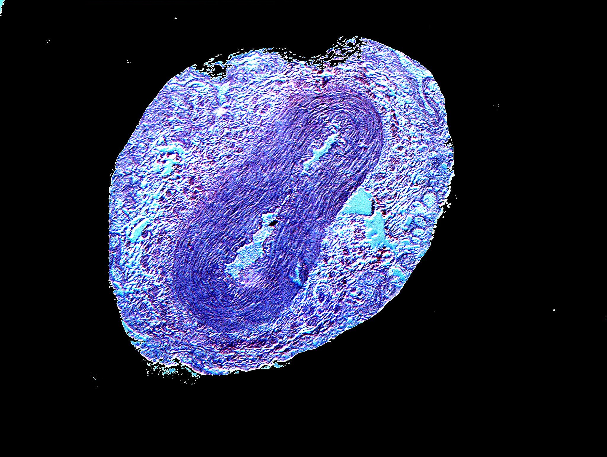
While we’re poking around on the inside of critters, we can take a look at a section of the vas deferens of a human. Now, if you were to ask “Vas the deferens”, Freud would give you a failing grade. This, of course is the duct that acts as a conduit for sperm to the urethra. The first image is more or less just a blob with virually no distinguishing content. However, with a bit of computer magic, we can manage to tease out some structure.
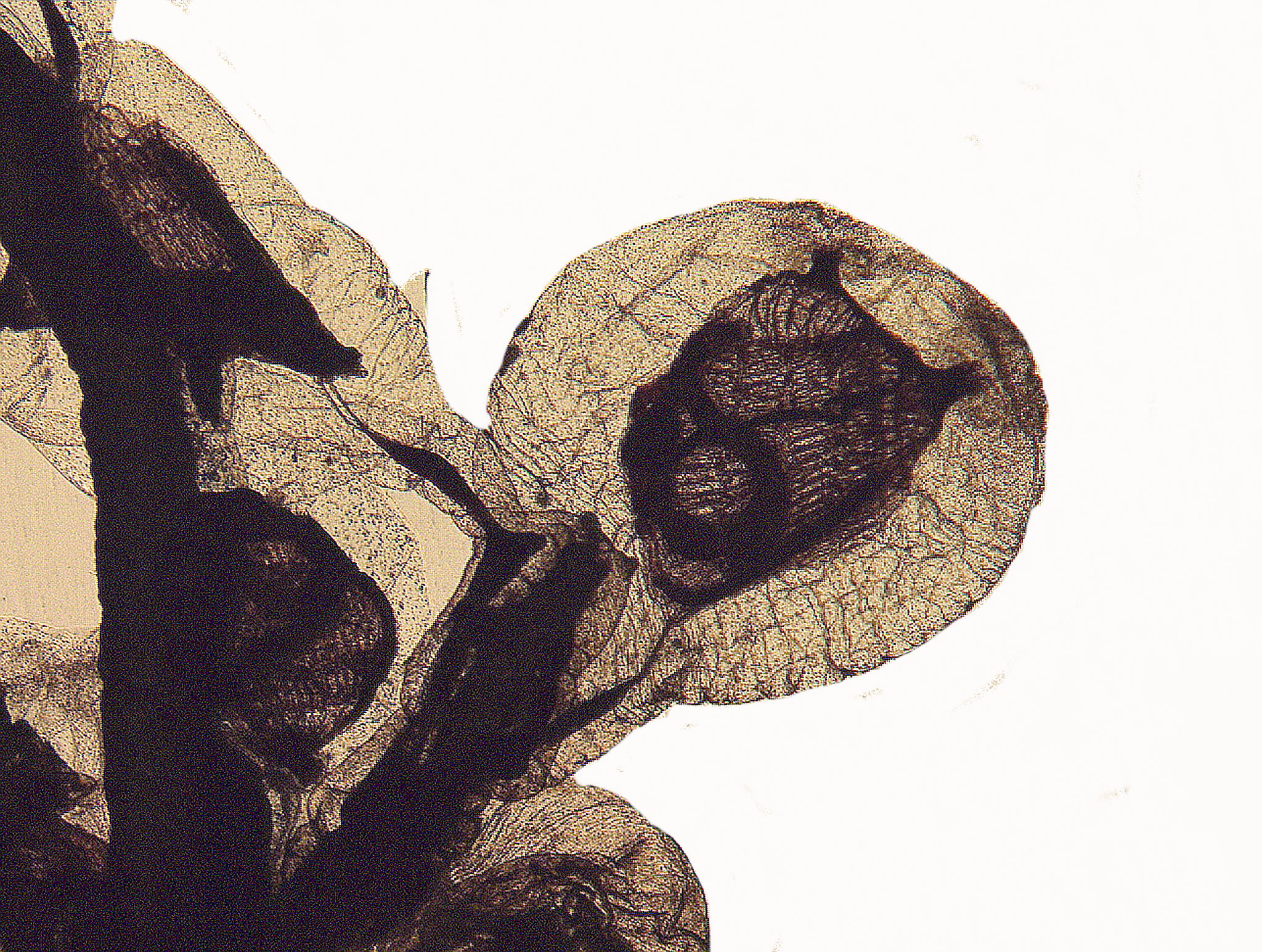
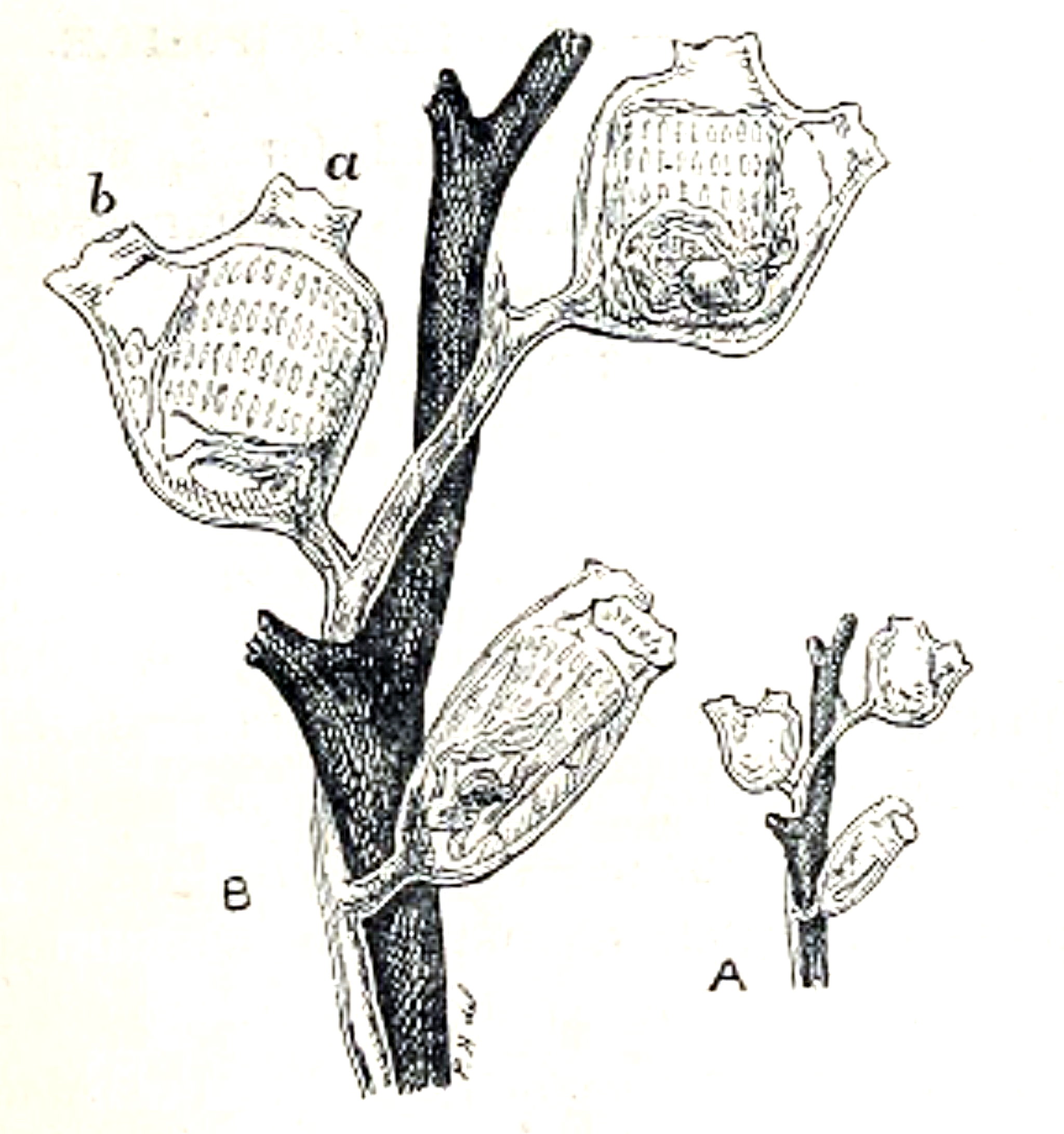
While sorting through these boxes of old slides, I came across one that especially caught my attention, a slide labeled “Zoophyte–Australia” and then there was a genus and species also handwritten. That information proved useless due no doubt to radical shifts in taxonomy. It looked very intriguing and, at first, I thought it was a hydroid. However, after I had taken several micrographs of it and processed them on the computer, I began to suspect that it was a colonial ascidian tunicate. I began a series of searches and finally, after nearly an hour, I found what I think is the most likely candidate, namely, a species of Pedophora.
First, I’ll show you a brighfield image, then an inverted one, and then an old drawing of Pedophora listeri. Remember that the specimen we are looking at is at least 100 years old and has undergone some shrinkage. The inverted image shows the siphons and the slits in the pharyngeal basket more clearly than the brightfield.

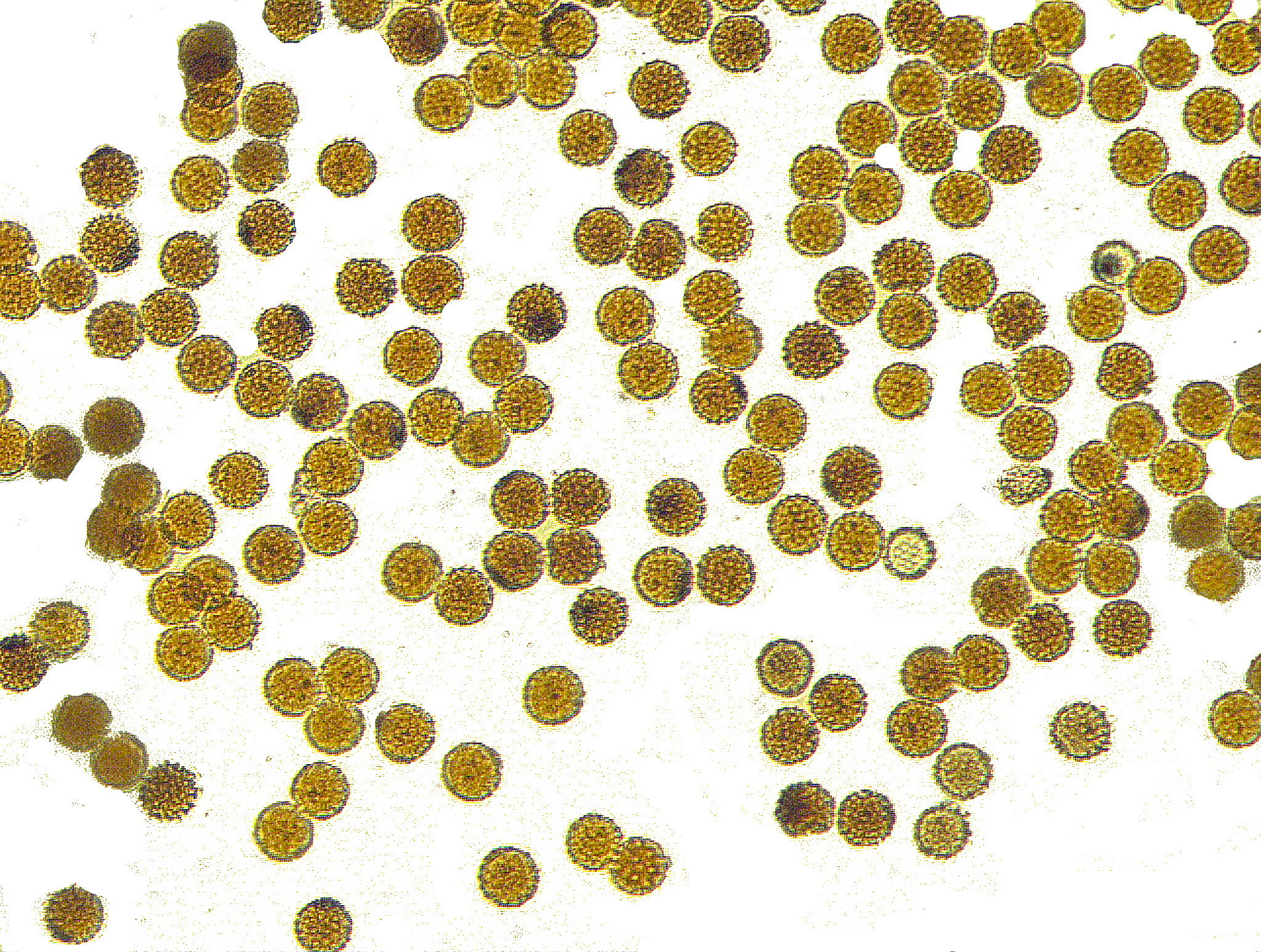
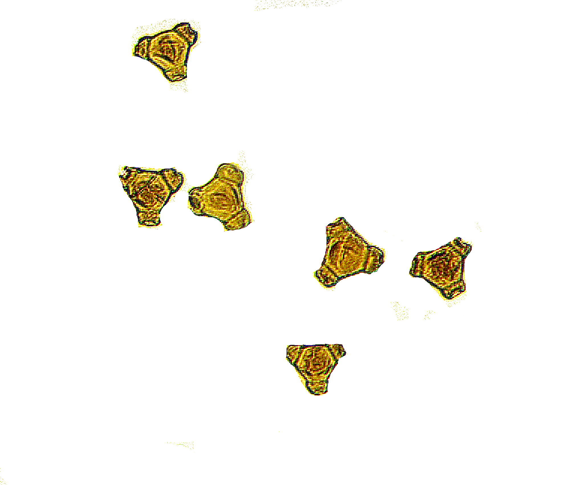
Finally, let’s conclude with images of 2 pollen strews. Neither are identified, but they are quite distinctive and quite different. The first consists of spiny, minute spheres and the second shows roughly triangular forms.
I hope you’ve enjoyed this little tour.