|
Rheinberg and Rainbows
Sawyer Winn, USA |
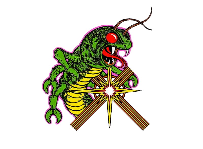
Video games have been something that I have always liked. The color and design of each game are very fun to look at and see how each game uses different combinations to give the game a different feeling to match the game’s story. One game that I found to be very interesting was the arcade game Centipede. I found this through my mother, who said it was her favorite game growing up. Centipede is about a forest of bugs that are trying to attack the player, but the color choice is unique for this game. Each level has a different color scheme, each time having two bright neon colors that contrast each other nicely. For example, the arcade game cabinet has artwork that I think shows the style of the game very well.

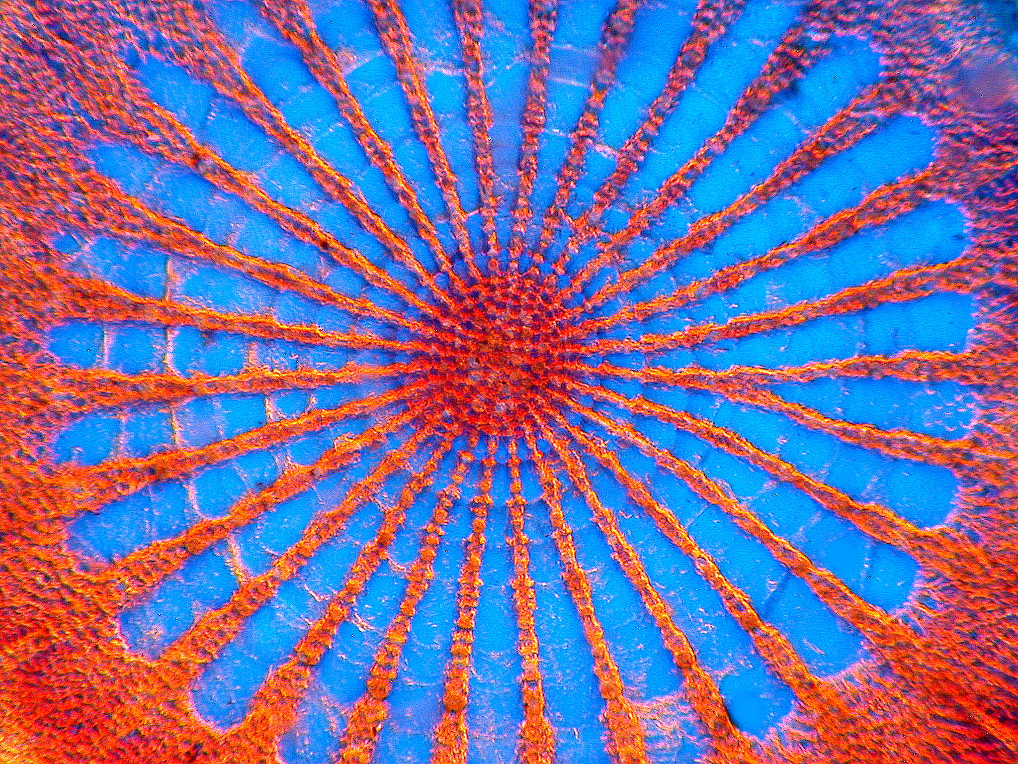
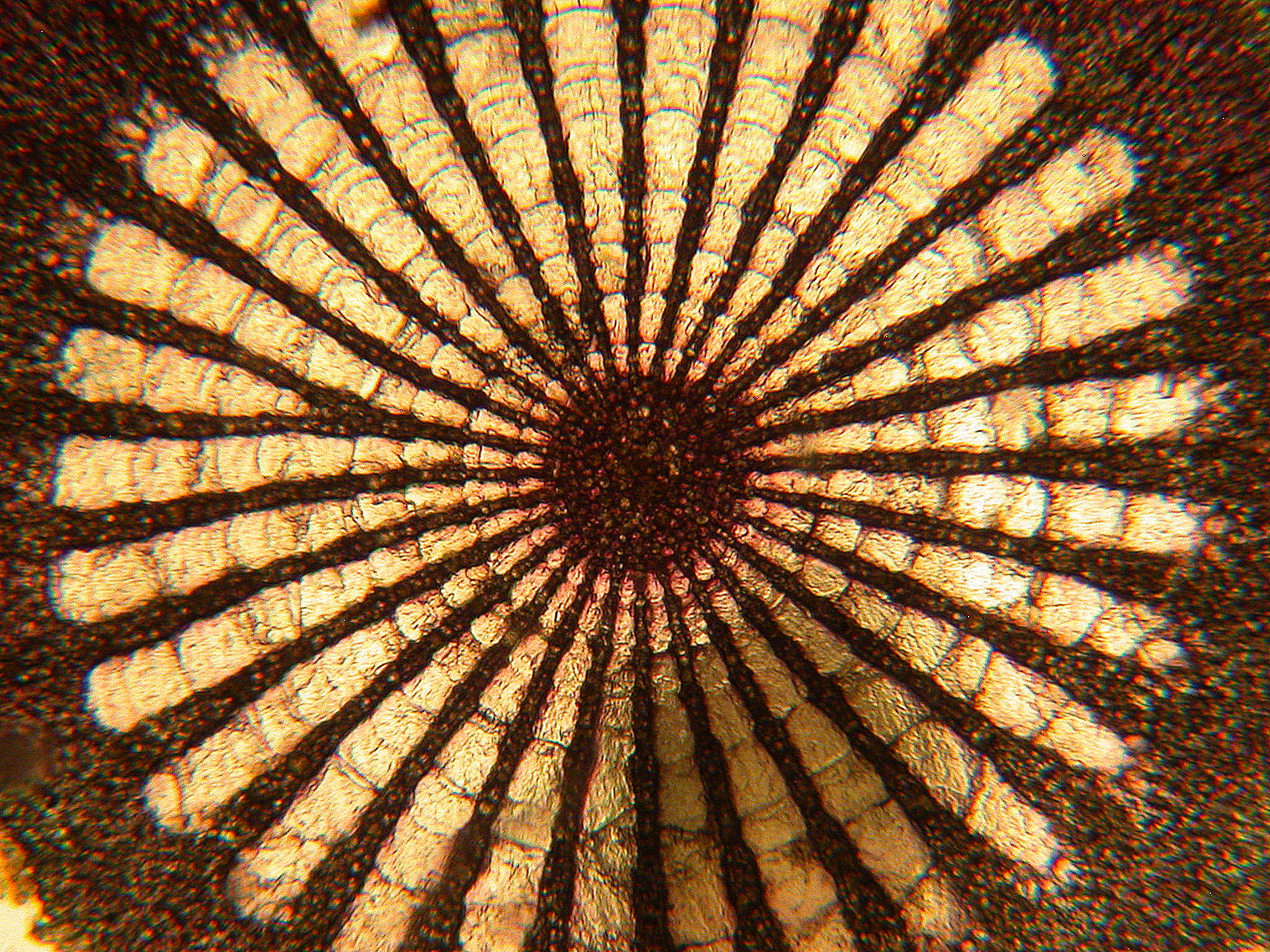
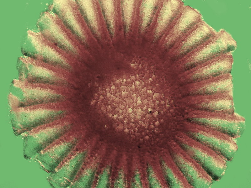
While photographing images on the microscope, I learned about a technique known as “Rheinberg Illumination”. This technique uses filters that have two parts, a ring around the edge that can be a solid color, or split into segments of different colors, and a smaller round filter in the middle of a contrasting color. When positioned correctly, the filters will cause the background to change to one of the colors, and the object being observed to be illuminated in another color. This can allow some beautiful patterns to be seen easier, and makes very colorful images that remind me of Centipede. This image is of a sea urchin spine cross-section, using an orange and blue Rheinberg filter. The image after that is the same spine, but without a filter, showing the difference the filter can make.
This one is a different sea urchin spine, but using the same filter. The thickness of the specimen can determine how much color comes through in the image. In this one, the structure of each spoke can be seen very clearly, with small crystals being tightly packed together along the spokes.
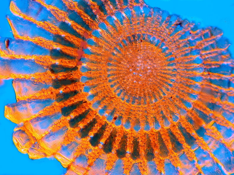
This is yet another urchin spine cross-section, but with a very different internal structure. The center is much wider than the previous two, and the spokes are closer together and wider. The filter was also changed, giving a different contrast to the specimen.
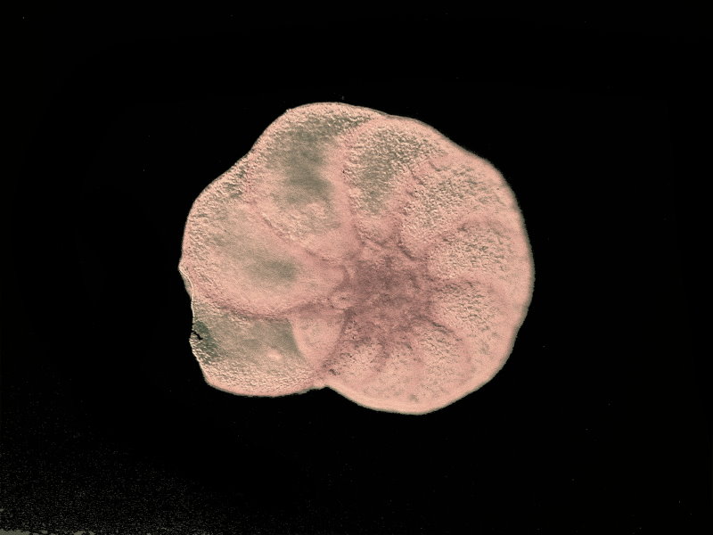
The next two are different forams photographed with the orange and blue filter and then edited to increase contrast. The small holes in the surface of the shells can be seen clearly, and the different sections around the spiral are evident in the first image, like that of a nautilus shell.
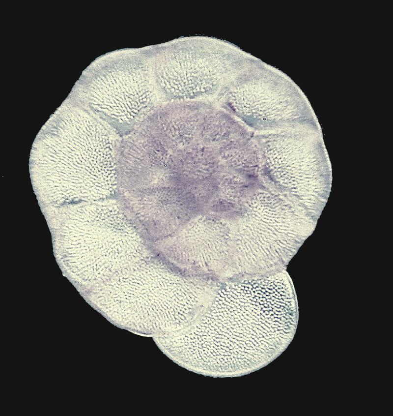
This is another shell-shaped foram, but with a green and orange filter. A different kind of structure can be seen on the surface. Instead of small holes like the others, this one also has needle-like grooves on it.
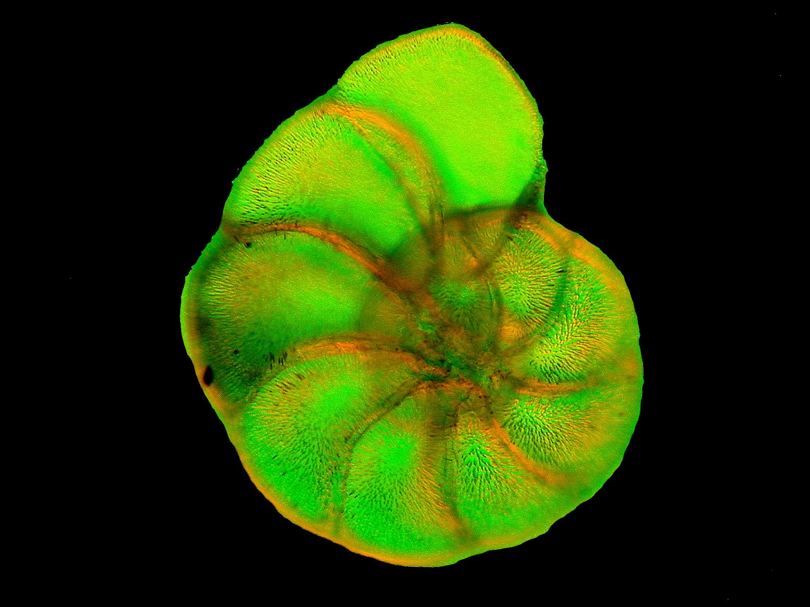
This one is a bit different, showing four different types of forams. Being single celled marine organisms, there are countless different types that exist. This leads to many, many pictures being taken that they can’t all be included in a single article. Each one has a different shape and texture, so it is really fun to explore all the types there are.
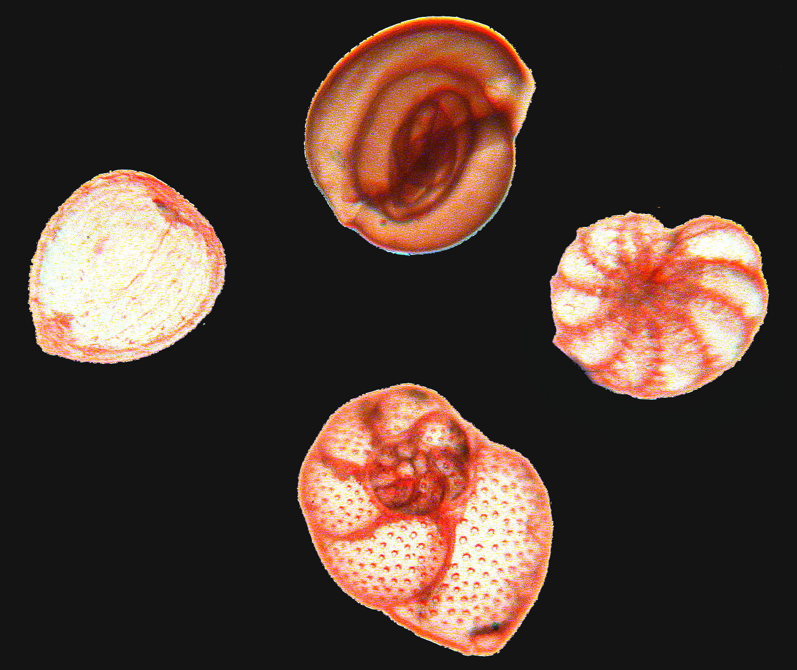
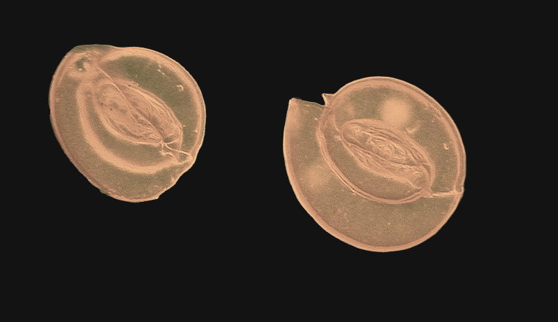
These are some odd shaped forams, with an oblong spiral shape. You can see how the spiral of these specimens gets tighter and tighter as it approaches the center, forming a football shape in the middle.
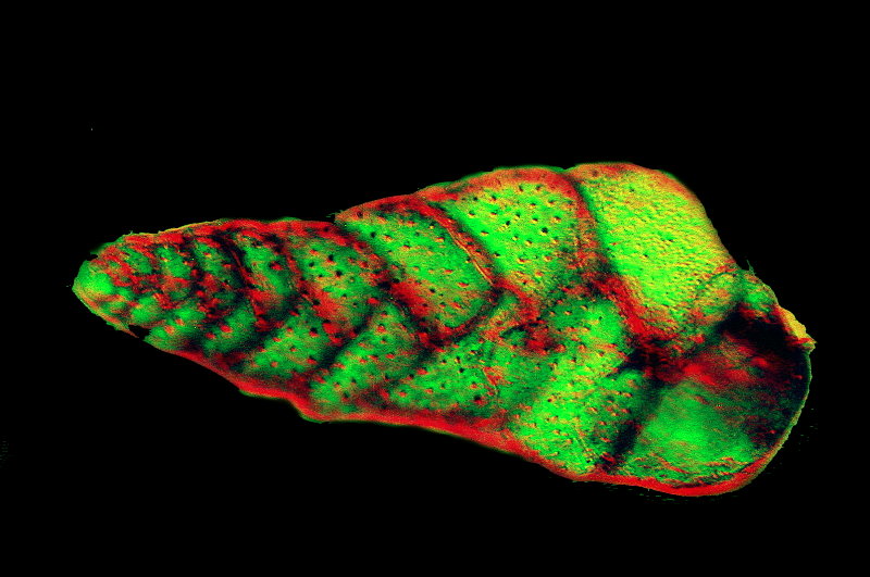
This is another foram with another different structure. It has a conch shell shape, and the internal chambers can be seen again, getting smaller as they approach the tip. The dimpled surface is also visible.
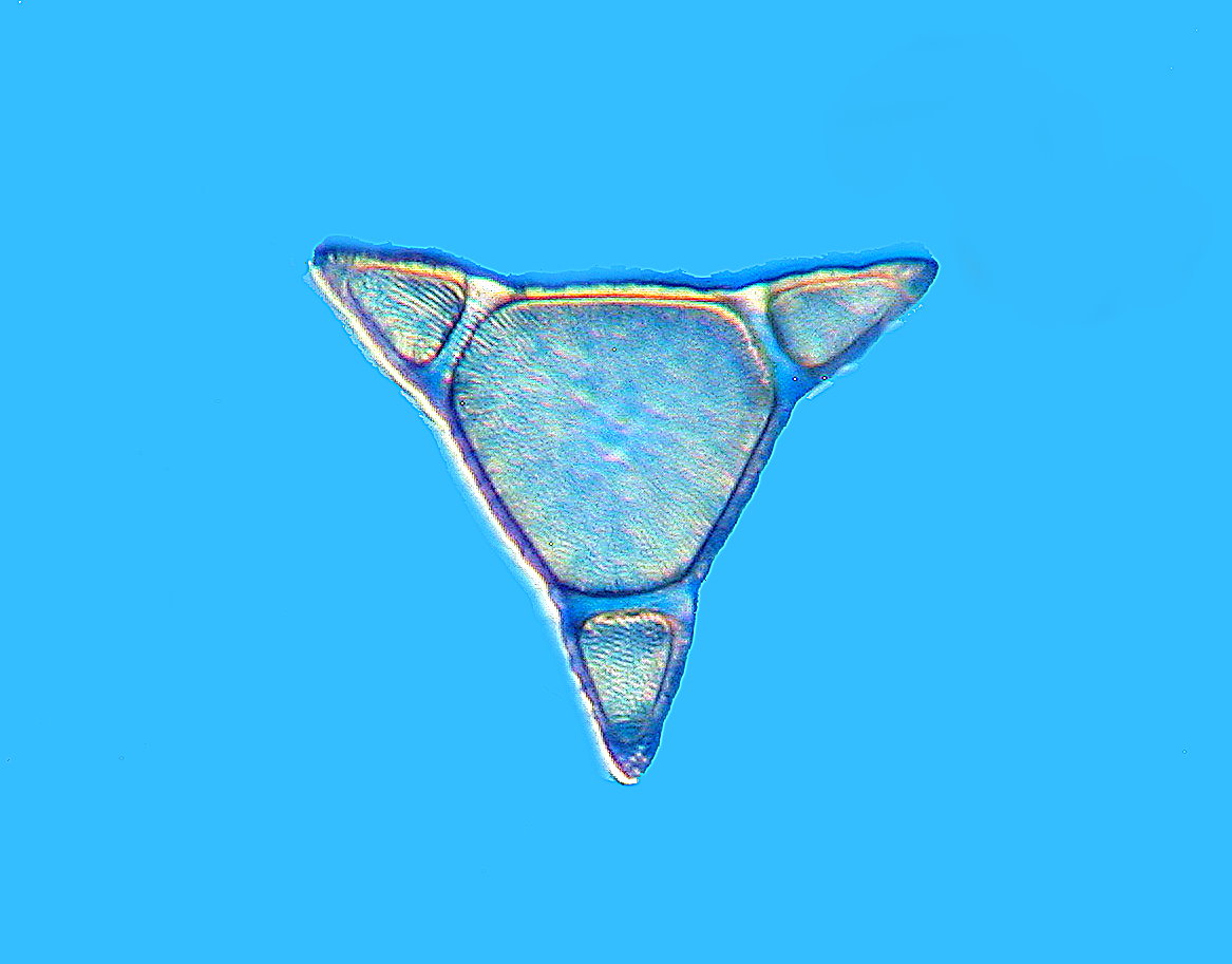
Last but not least, this is a diatom. Diatoms are types of algae that have a shell-like structure. This one is called Triceratium. It forms a prominent triangular shape. This filter used a blue filter with no center ring, making the diatom look white when photographed.
All of these images show how unique these small organisms can be. Using Rheinberg Illumination, you can enhance the structures in each specimen, creating beautiful colors in the process. Because of the neon colors in these, they look like a retro video game arcade with bright lights and heavy saturation. This is one of the reasons why microscopy is so fun, because you can make the images look however you want, while still showcasing the fantastic specimens you observe.
All comments to the author Sawyer Winn via Richard Howey are welcomed.
If email software is not linked to a browser, right click above link and use the copy email address feature to manually transfer.
Microscopy UK Front
Page
Micscape
Magazine
Article
Library
Published in the June 2022 edition of Micscape Magazine.
Please report any Web problems or offer general comments to the Micscape Editor .
Micscape is the on-line monthly magazine of the Microscopy UK website at Microscopy-UK .
©
Onview.net Ltd, Microscopy-UK, and all contributors 1995
onwards. All rights reserved.
Main site is at
www.microscopy-uk.org.uk .