Ringlights are very useful for 'fit and forget' incident lighting on stereo microscopes. More recent models tend to use one or more rings of LEDs, although the models based on either fluorescent tubes or fibre optic light sources are also available. I use the YK-B144T model with 144 LEDs which was reviewed in the July 2012 issue of Micscape. The ten intensity settings and ability to control the four light quadrants independently allows useful modelling when desired.
I hadn't given much thought to the potential alternative uses of ringlights until read a message in the Yahoo 'Microscope' Forum by Ricardo Tsukamoto where he highlighted and linked to a paper by K F Webb entitled 'Condenser-free contrast methods for transmitted light microscopy' (JRMS, 2015, vol. 257 (1), 8-22). The paper described the use of fabricated rings of LEDs for various compound microscopy techniques without the need of a condenser. Techniques illustrated included phase, darkfield and Rheinberg. The paper made me wonder how well the ringlight worked as a darkfield condenser on my stereo as I didn't currently have a ready way of doing this. With a little background reading, I wondered why I'd never tried this before as Brian Bracegirdle in his invaluable book Scientific PhotoMACROgraphy (1995) describes and illustrates the successful use of fluorescent and fibre optic ringlights for darkfield macroscopy studies.
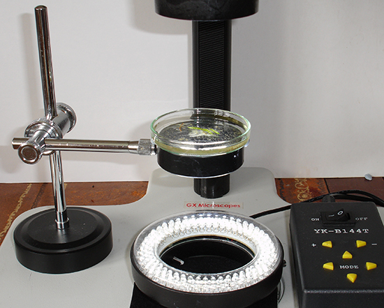
So without further ado, I tried the 144 LED ringlight for darkfield on my Leica S8 stereo. The three concentric rings of LEDs are aligned to face inward to provide a bright central spot of light at the typical working distance range of a stereo of ca. 80 - 100 mm. The sample therefore ideally needs to be at least 80 mm from the ringlight. The setup I tried is shown below.

Above. Darkfield setup on a stereo microscope. The control box of the 144
LED ringlight offers ten intensity settings and with each quadrant independently
switchable. A piece of black felt sits under the ringlight for a black
background because little if any LED light reaches it. A magnifying glass stand
with lens removed was adopted as the support and suited the 60 mm diameter Petri
dish. Its compact form and adjustability for height and angles made it ideal.
The sample can be scanned by moving the stand rather than the dish. The dish can be immobilised with a little Plasticine or a larger annular ring added as additional support.
The setup gives an excellent velvety black darkfield for the full range of optical settings of the 1-8X zoom on the stereo. The LEDs are very bright and only set near their minimum intensity at lower mags. For photography it's best to set a level sufficient for good darkfield but without excess glare. Because the LEDs face inwards, I don't find any shrouding is required to reduce visual glare while at the stereo although card baffles could be readily added.

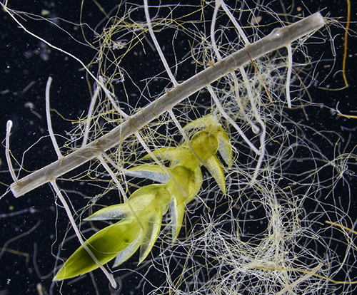
Above at 1X zoom (10X optical mag). Darkfield is very useful for spotting small organisms, either planktonic or epiphytic, for isolation to study under the compound. Also a very pleasing way to study the larger life in situ under less stressed conditions for the fauna than a cell under a compound microscope may give. LEDs are a cold light source as well.
I'm delighted with this additional darkfield feature for my stereo studies at no extra cost. Live pond life in particular is striking. Samples, especially in water, are more vulnerable supported above the stereo base but with care works well. A stereo with a generous focussing pillar is also needed to focus at the higher focal plane. A seat with adjustable height or a deep cushion ready to hand is also needed to maintain a comfortable posture at the higher eyepiece level.

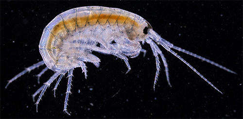
Above. Freshwater shrimp, (Crangonyx pseudogracilis?) optical mag 25X. The shrimp was very active so ISO 1600 EV-2.0 was used (Sony NEX-5N body, IR handheld shutter remote). No post capture adjustment was made to the black level in the image.

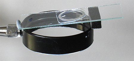
Above. This admittedly rather precarious temporary setup can be useful to maximise the plane of focus for isolated larger specimens photographed under a stereo (e.g. the shrimp above). The sample on a ringed slide with coverslip is raised one end so angled at ca. 6º; i.e. perpendicular to the optical axis of the tube used for photography.
Compact darkfield condensers for stereos using existing base lighting are readily available—Asian based eBay sellers offer one design at typically ca. £60+ incl. postage. I've never tried one as they typically cost more than my ringlight and may require high quality stereo base lighting to work well. They also don't have the ready control of the lighting direction and intensity which the ringlight offers. (With Micscape Editor's hat on, we would welcome readers sharing their experiences on how well these other designs work, or other methods readers use for darkfield stereo work. One or more flexible light guides directed from below and upwards at an angle to the subject is one approach.)
The YK-B144T ringlight also works very well as a low power darkfield 'condenser' on my Zeiss Photomicroscope III without a condenser or auxiliary lens present—creating velvety black full field backgrounds for objectives from 2.5X up to 10X NA0.22. At the moment a Zeiss achromatic-aplanatic condenser with a darkfield 'D' setting with top condenser lens removed works very well in this role (see article in the October 2008 issue of Micscape). But the ability of the LED ringlight to control quadrants may offer possibilities to 'model' subjects under darkfield and something to explore in the future.
Comments to the author David Walker are welcomed.
Acknowledgement. Thank you to Ricardo Y Tsukamoto for highlighting the paper by
K F Webb in the Yahoo Microscope Forum, May 22nd 2016.