|
Using the 'Mark One
Eyeball' Written by David Walker |
|
Using the 'Mark One
Eyeball' Written by David Walker |
| I was watching a TV
documentary recently and a pilot was being interviewed on
the flight deck of a large jet surrounded by a plethora
of instruments and high tech gadgetry. The pilot was
asked which was the most important instrument in the
cockpit. Without a pause for thought he pointed at his
eye and replied, "the Mark One Eyeball, by far the most
valuable instrument since the days of earliest flight." Perhaps this is a message that, as enthusiasts of microscopy and the close-up world, we could well remember. Despite all the marvellous advances in microscopy and the many gadgets both optical and electronic to help study the world around us, the importance of the Mark One Eyeball sometimes gets overlooked. We couldn't, of course, use any of these instruments without our eyes, but we can also enjoy much more of nature in miniature with no optical aid at all ... by just using that all important Mark One Eyeball ... but we need to get up close! Try this simple experiment. You are probably over 50 cm from your computer display at this moment and unlikely to see the fine structure of the screen, (i.e. phosphor or individual LCD pixels). Now move to whatever is your closest comfortable viewing distance, but still able to focus your eyes. At this distance you'll probably start to see some screen structure*. Try the same thing with the fabric of the clothes you are wearing, when the weave of finer fabrics will be seen. [*Ed's note: not so easy 13 years since first writing with the now common higher resolution screens, especially the Apple Retina series.] Most of the optical instruments to help us study the macro and microscopic world have one fundamental role - to present a larger image of the subject to the eye. This is effectively what you do when you get up close to something to appreciate the finer detail, whether reading small print, threading a needle, etc., or as above, getting closer to the computer screen. "Heh David, aren't you stating the obvious?" Perhaps I am, but how often, when we enjoy the countryside or our gardens, do we get up this close to nature? I know I don't as often as I should. In Beyond magazine and its sister magazines, readers are encouraged to use a hand lens, (ca. 5x to 10x), as a simple optical aid while out on a walk. But even without this, if we get into the habit of getting up close when we see something of interest, whether its a tiny flower, lichen, or moss on a wall, there's an amazing wealth of extra detail that could be revealed.
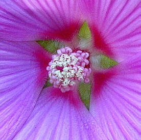
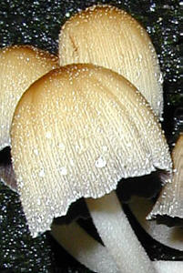
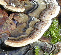
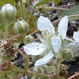
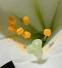
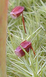
In my back garden there are various small plants that come into bloom each year and I usually just admire them standing up and rarely from a close viewing distance. But I noticed one tiny plant stem was sticky to the touch, so I held the stem up close, viewed it against the light, and was surprised to see wonderful stem hairs with red glands on the tips. Taking a piece of the plant indoors to view under the stereo 'scope gave a better view, but it's this first step of getting closer that can give us a new insight into many of nature's wonders. So when you are in your garden, out walking, or on holiday, stop awhile and get really close to features in nature that attract the eye. You may be surprised at just what that Mark One Eyeball can reveal. Have fun! A selection of subjects that attracted the author's eye near his home in northern England are shown below and above. These are mainly plant subjects but studying e.g. insects up close can be just as rewarding.
|
|
Comments to the author David
Walker welcomed.
All images by David and Ian Walker using a Nikon Coolpix 700 in
macro mode.
First published in the March 2000 edition of the e-Zine 'Beyond' edited by the late Marly Cain-Fryman.
Acknowledgement: Thank you to Jessica Cain for copies of the author's 'Beyond' articles. The original 'Beyond' site is now sadly closed.
Re-published in the June 2013 edition of Micscape. Please report any Web problems or offer general comments to the Micscape Editor .
Micscape is the on-line monthly magazine of the Microscopy UK web site at Microscopy-UK
©
Onview.net Ltd, Microscopy-UK, and all contributors 1995
onwards. All rights reserved.
Main site is at
www.microscopy-uk.org.uk.