|
The Quest for Spicules Part 2 by Richard L. Howey, Wyoming, USA |
In this part, I want to focus primarily on skeletal structures in sponges and soft corals (Gorgonians). As I mentioned before, there are 3 primary types in sponges in terms of composition: calcareous, siliceous, and organic (spongin) and I want to look at some examples of all 3 plus some calcareous Gorgonian spicules. You will notice that a number of the images here appeared in Part 1, but I am repeating them here to raise certain issues and also to emphasize the character or some of these structures.
There is an almost unavoidable tendency to write, talk, and think about non-human organisms in anthropomorphic terms which points to a deeply embedded sort of linguistic and conceptual Urgrammatik. I mention this because I was just about to assert that sponges are highly opportunistic, but opportunism is a human trait which some people, especially politicians and other greedy sorts, have made a specialization. What I really want to convey is that sponges have an extraordinary ability to adapt and thrive in a wide variety of ecological niches, some of which are rather surprising.
If you have ever collected shells out of tide pools, you may well have noticed small, thin layers of sponge, bryozoa, etc. growing on them. Also, if you have been out beachcombing, you have very likely found shells with small round holes in them, so neatly done that they look like they have been drilled. Well, that’s almost the case; these holes were produced by some type of very interesting boring sponge. These sponges have a number of deeply weird characteristics: 1) They begin their attack on shells while they are still in the larval stage, 2) There are special cells which are amoebocytes that remove tiny chips of the calcareous material of which the shell is comprised, and 3) the sponges then extend themselves down into these tiny caverns and continue to grow and expand their boring activity, which I have already pointed out is really quite interesting. Remember that sponges are bizarrely colonial organisms and boring sponges can spread themselves in thin sheets to accommodate virtually any shape whereas other species are programed to produce a distinctive, patterned structure. The former is a significant part of what I meant when I earlier referred to sponges as opportunistic. Take a look at how a sponge has accommodated itself to a quite thorny sea urchin spine.

In this case, the sponge growth is such that it looks like a kind of webbing between the barbs of the spine.
In contrast, we have those sponges which are genetically designed to produce quite consistently identifiable architecture. Euplectella, the beautiful Venus Flower Basket, produces these splendid tubular structures.
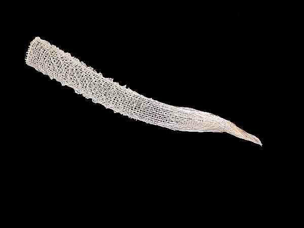
When examined closely, one can observe the lovely, intricate lattice work which constitutes this skeleton.
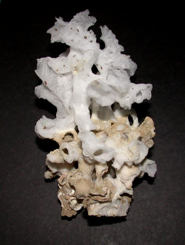
Another intriguing mystery is that certain types of sponges tend to be associated with a high degree of regularity with other organisms. For example, there is a mollusk, Xenophora, which for some inexplicable reason tends to attract a variety of opportunistic hitchhikers including a rather distinctive glass sponge, an example of which you can see here still attached to the shell.
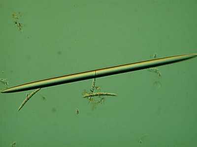
The forms of sponge spicules can vary enormously as I mentioned previously. Earlier, I showed you some monaxons from marine sponges. This time, I’ll show you an example from Spongilla, a freshwater sponge.
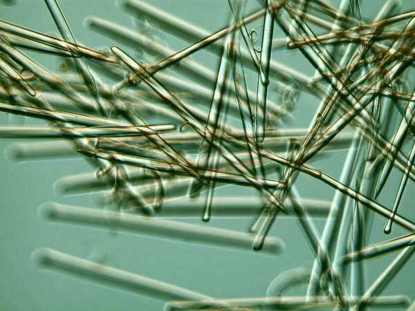
In marine forms, monaxons can occur in very high densities.
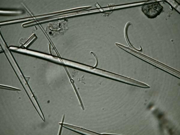
However, even in sponges where the spicules are predominately monaxons, one can find other forms, such as the “C” shaped forms in the image below.
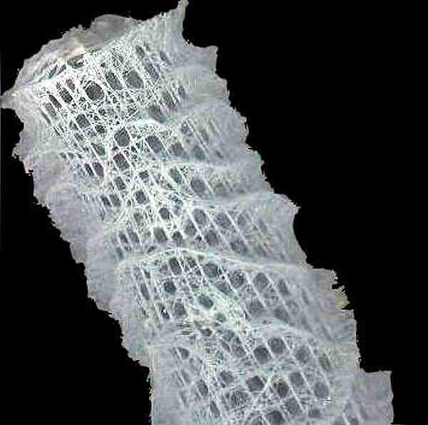
In Euplectella, we saw an example of an elegant form which sponges can produce and typically glass sponges form intricate lattices of remarkable complexity and beauty. Here is a piece of such a lattice.
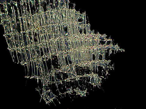
And here is a close up to give you an idea of the intricate networking of the spicules.
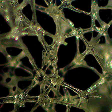
Sponges, like corals, produce a staggering variety of forms. There are sponges that look like elegant vases in an incredible range of colors; others that look like a cluster of fingers; and yet others, like some of the glass sponges, that look like some futuristic, and perhaps slightly inebriated, architect designed them. However, many other sponges easily go unnoticed and seem quite undistinguished. A rather typical such example can be seen below.
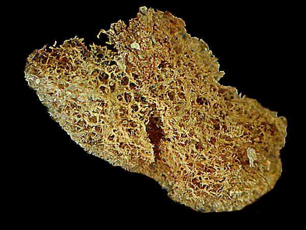
Often, however, our tendency to gloss over or dismiss a particular organism as “uninteresting’ is a consequence of our not having looked carefully and closely enough. Consider this close up of a bit of sponge.
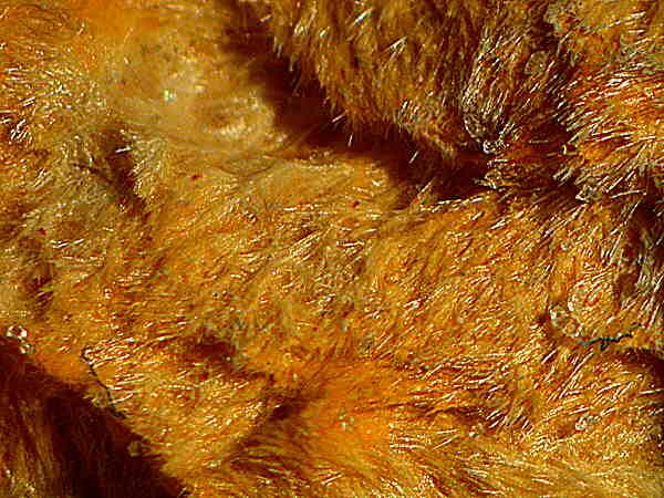
At first glace, it appears to be just a yellowish-brown clump of nothing at all interesting. However, if you take a second, more careful look you will see that this clump is a mass of acicular (needle-like) spicules which form an amorphous, but very complex, skeleton.
From amorphous, let’s proceed to a sunburst. I found this splendid little specimen on a spine of a tropical sea urchin.
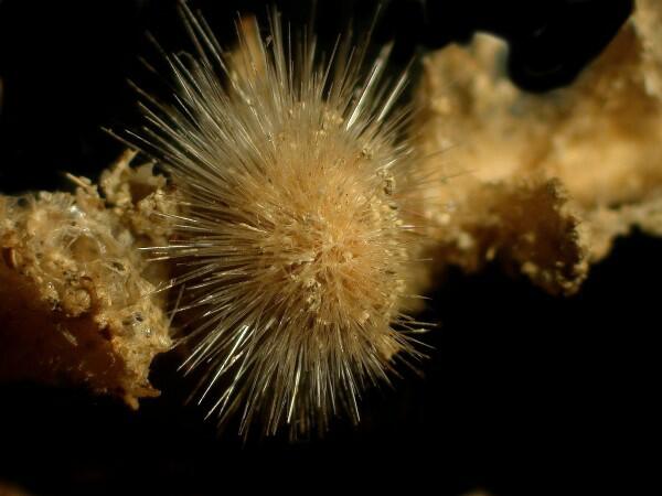
Interestingly, individual spicules show almost as much variation as sponges themselves. Consider this “broadsword” spicule from a glass sponge.

There’s a great name for some future rock group: “The Dueling Broadsword Sponges”.
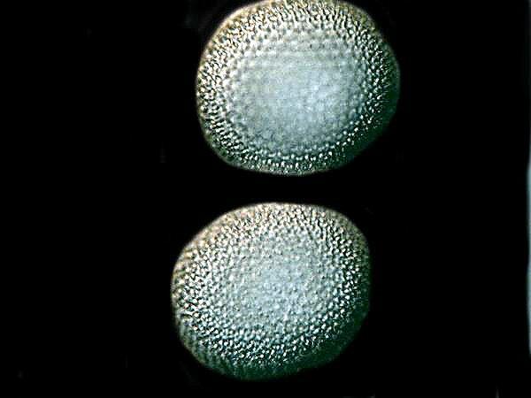
Now take a look at these wonderful, spiky slight oblate spheroid spicules which come from a sponge which at first glance looks like a piece of coral
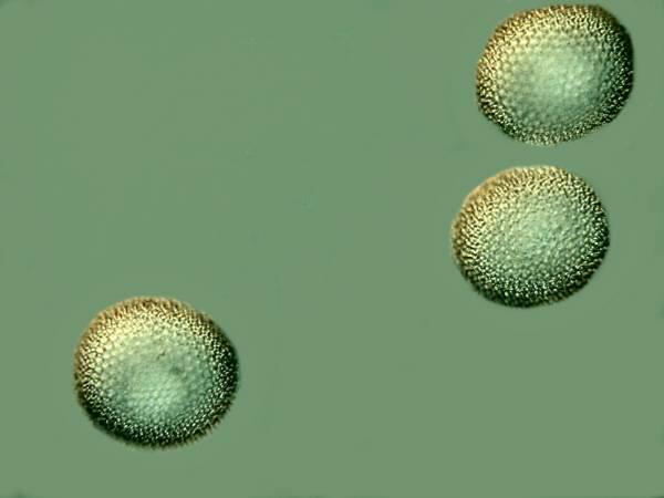
And here’s a closeup.
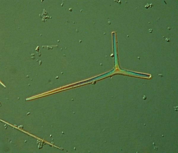
In addition, there are triaxon spicules which, in this case, turn out to be nicely birefringent.
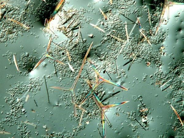
And a closeup:
Also, there are the spectacular (or should I say, spictacular?) spicules which one can find in certain hexactinellids (glass sponges). The one below comes from a specimen of the genus Hyalonema.
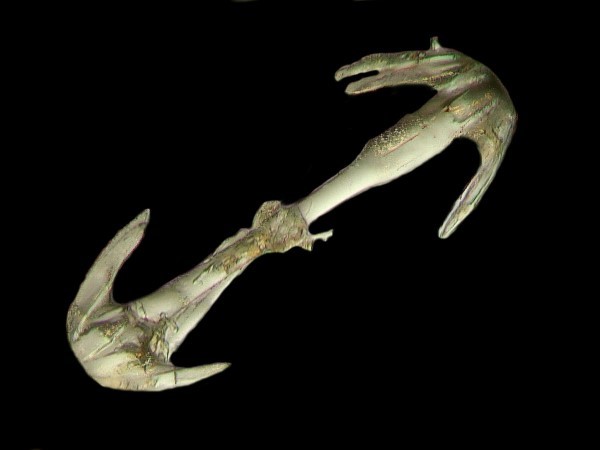
This sponge is one of the strangest I have ever encountered and I intend to devote an entire article to it in the very near future.
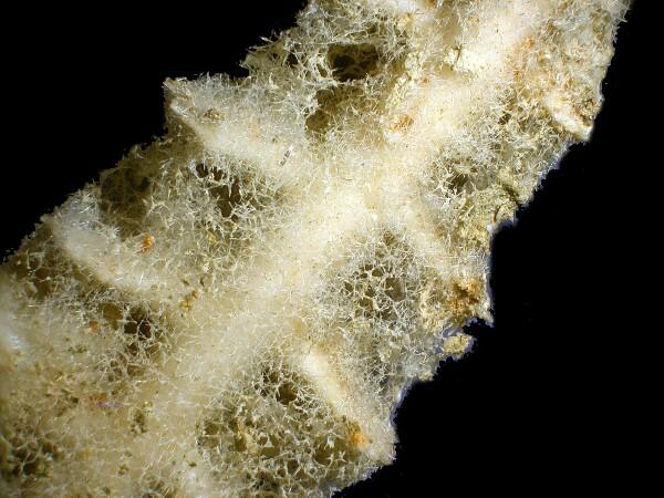
Since we have looked at some examples of calcareous and siliceous spicules, let’s now take a brief look at an example of a sponge skeleton composed of organic material, namely, spongin. In this particular type, it appears as a net-like mesh which you can see below.
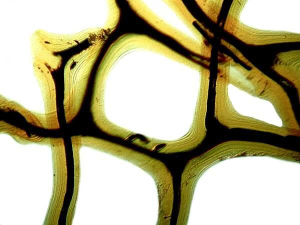
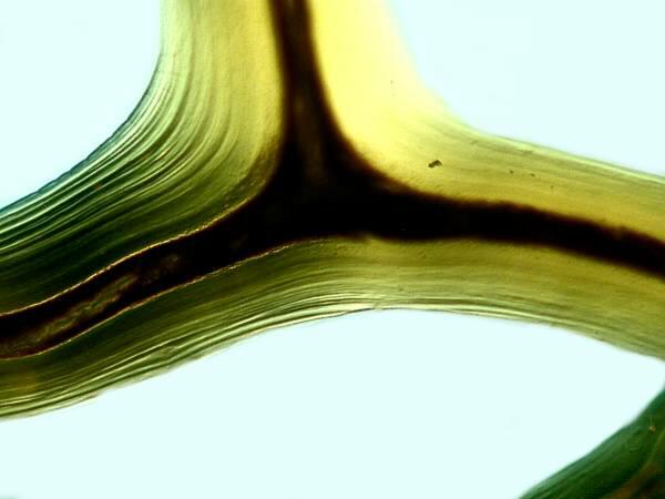
The darker central area is the core around which amoebocytes deposit layer after layer of the lighter-colored yellowish organic material. These layers are clearly visible in the image below.
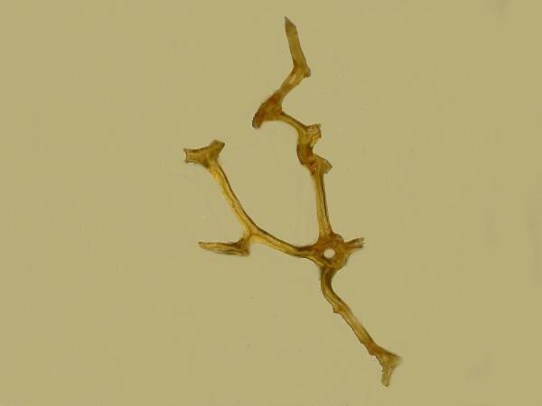
I happened across a fragment of this skeleton which to me suggests the figure of some tiny alien mannikin performing an exuberant dance.
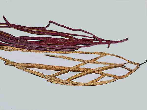
Finally, let’s take a look at some spicules from some soft coral. Soft corals come in a rather wide variety of shapes and sizes and certain types are popularly known as “sea whips” or “sea fans’; these are the Gorgonians. Below is an image showing portions of a red one and an orange one from Florida.
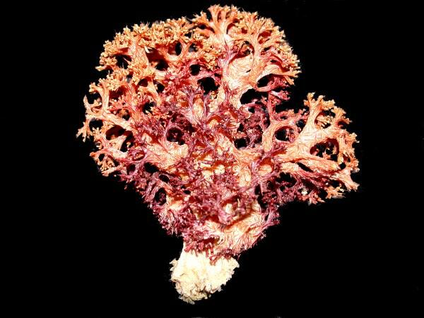
Here is an example of a bushy soft coral from the Philippines.
If we take samples of the orange and red Gorgonians and place them in bleach to dissolve away the tissue, we discover 3 remarkable facts: 1) The spicules occur in enormous numbers, 2) They are morphologically quite distinctive, and 3) They retain their pigmentation even through bleach, alcohol, and formaldehyde as you can see in the 2 images below; the top one being spicules from the orange one and the second one, from the red one.
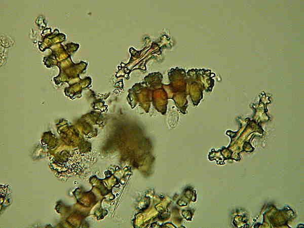
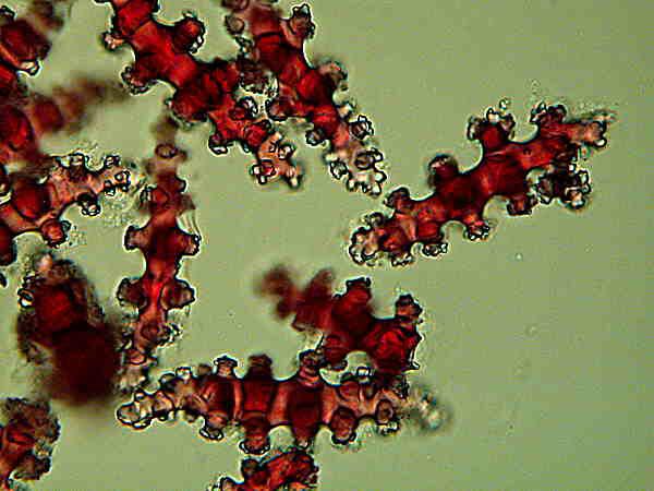
The third fact is worth taking note of, since many pigments in marine invertebrates disappear very quickly after preservation. In this case, it seems that the pigments are bonded into the crystals in the calcareous material as the amoebocytes deposit them in the formation of these skeletal structures so that they remain more or less impervious to the ravages of chemical attacks.
Skeletal structures in invertebrates continue to be a source of great fascination to me and so you can expect more ramblings from me about these remarkable entities in the future.
All comments to the author Richard Howey are welcomed.
Editor's note: Visit Richard Howey's new website at http://rhowey.googlepages.com/home where he plans to share aspects of his wide interests.
Microscopy UK Front
Page
Micscape
Magazine
Article
Library
Please report any Web problems or offer general comments to the Micscape Editor .
Micscape is the on-line monthly magazine of the Microscopy UK website at Microscopy-UK .