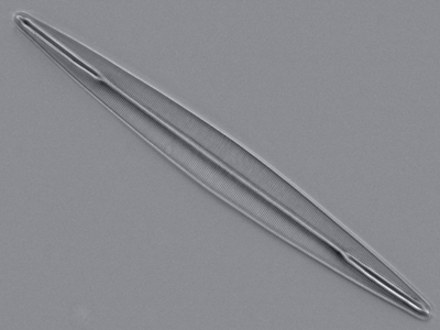
Editor's note: The Editor first came across Osamu's work from a reference in Rene van Wezel's recent article Diatoms and microscopy, a contrasting combination . Later visiting Osamu's website, the images of test diatoms, notably A. pellucida were some of the finest I recall seeing in visible light. Osamu shares aspects of his studies below.
Osamu writes: I started diatom frustule observation in 2001 as a hobby using a research grade microscope. I thought that optics knowledge is VERY important for understanding microscopy when I looked at diatom frustules under microscope. I also started to study optics as a hobby. Thus my paper describing illumination is no relation to my research field! But I like the world under the microscope (also telescopes) using visible light.
Editor's note: Osamu summarises his techniques as follows.
Method overview for A. pellucida
imaging:
Objectives: more than 1.2NA. Usually I use Nikon PlanApo 60x (1.4NA) and
Olympus Ach (for pol) 100x (1.3NA).
Condenser: achromatic aplanat
(1.4NA) or Darkfield (1.2-1.43NA)
Wavelength of illumination: 440-500nm.
Green filter (546nm) is not suitable.
Light source: Halogen 100W
lamp with IF filter (450-500nm).
Blue LED (lumileds, 470nm) or white LED +
IF filter are also adequate.
Illumination method: essentially col (annular
oblique) + pol.
Digital
camera: Nikon Coolpix 990 and 995
Mountant: pleurax (Wako Pure Chemical
Industries, Ltd.)
In the condition of high NA (>1.0) image contrast is
very under low normal lighting. Polarized light is important for high NA studies.
Resolving the punctae of A. pellucida is not difficult if you use oblique +
polarized light.
Slide preparation: Usually I use Pleurax (RI=1.7) for a diatom mountant and best coverslip, but have used Realgar in the past.

A.
pellucida, Nikon Coolpix 995, single image of whole diatom. Image needs
to be clicked to view punctae detail on the master.
A Nikon Coolpix 995 with 10x eyepiece can record the full length of A.pellucida and punctae at the same time. Please see above image.
The large image (941 * 5570) above, click to view, is the result of a combination of about 7 individual 1280x1024 monochrome CCD pictures.
To minimise slight exposure differences and the slight tonal changes which can make seamless merging tricky, Osamu remarks: Background subtraction is highly recommended. LED lighting with stable power supply is suitable for image composition. Light intensity from halogen and/or tungsten bulb is always fluctuating because of variation of power supply and lamp temperature.
Using polarised light and annular oblique for imaging
Osamu's paper can be downloaded here: A new method for visualization of diatom striae by using annular illumination and polarized light. Bull. Plankton Soc. Japan 51(1): 25-33. The paper is in Japanese but the extensive abstract and image captions are in English.
In addition to Osamu's work reported in his paper, Rene van Wezel's Micscape April 2009 article reporting of his own studies and citing Osamu's paper shows very effectively the benefit of annular oblique with polarised light for imaging punctae of A. pellucida.
Osamu's images of other test diatoms are on this page of his Microworld Services website.
Editor's footnote: The use of polarised light to aid diatom resolution has been reported by other workers and the Editor / author would be interested to learn of the earliest studies, especially if used with axial or annular oblique lighting. Google Book searches of online old texts and journals have been inconclusive, partly due to the varied way of referring to oblique and / or annular lighting.
