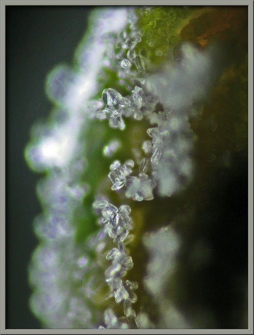
A Close-up View of the Hybrid

Delphinium  'Guardian Blue'


Delphinium elatum 'Guardian Blue'


by Brian Johnston   (Canada)


Over the past few years, I have searched in vain for an exceptional Delphinium to photograph for a Micscape article.  Finally, during a recent trip to my local greenhouse I noticed, behind a large grouping of white examples, this single, extremely colourful plant.  My search was finally over!

The cultivated Delphinium, (a member of the Ranunculaceae, or Buttercup family), possesses long, showy racemes containing variously coloured spurred flowers.  (A raceme is an unbranched, elongated flower cluster in which each flower is attached by its stalk directly to a central stem.)  Delphiniums are often referred to as Larkspurs, although this appellation  is also used by the related genus Consolida.  The genus name Delphinium derives from the Greek delphinion which refers to the dolphin–like shape of the bud.

Delphinium elatum ‘Guardian Blue’ possesses the strikingly colourful flowers seen above.  The small central petals are white, and they are surrounded by rings of larger, patterned, purple petals.  A whorl of mottled blue and purple petals provides the background.

The 25 centimeter long flowerhead begins to bloom at the base of the raceme, and about a week is required for the process to be completed.  The overall height of the plant is about one metre.


A closer look at the unopened buds reveals that they do rather look like fat dolphins (or tadpoles), except of course for their colour!  Notice that a spike-like green leaflet grows from the point of connection of each bud’s stalk to the stem (axil).


The two views that follow show an unopened bud with its spur (left), and a later stage bud in the process of opening (right).


Mature flowers are about 3 centimetres in diameter, and are usually so tightly packed in the raceme that they obscure the plant stem completely.  Note, at the top of the two images, the two smaller blooms that are in the process of opening.


These two flowers can be seen more clearly below.


One of the main reasons for choosing this particular hybrid is the intricate colour pattern possessed by its petals.  The image at right below, shows that this pattern is reminiscent of a stained-glass window.


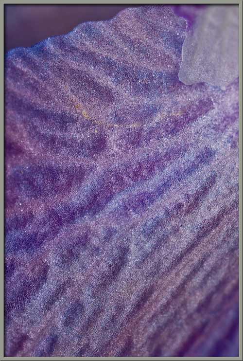
If we use the microscope to take a closer look at one of the blue petals from a flower’s back whorl, it is obvious that its surface is very three-dimensional.  Near the centre of the right-hand image there is a very tiny, pearl-like sphere on the end of a fragile stalk.  I have no idea of the purpose of this structure.


Near the tip of a similar, less mature petal, there is an area with greenish colouration.


Photomicrographs showing the pigmented cells within this section of petal appear below.


A higher magnification photomicrograph of the cellular structure is again reminiscent of a stained-glass window.


Between the green and blue areas of still another petal, there exists a pinkish colouration.


Low and higher magnification images of the cells in this area can be seen below.  The image on the right hints that the entire cell is not coloured.  Instead, there appears to be a thread-like internal structure which possesses colour.


Now let’s look at one of the large pink petals from the front of a flower.  Again, the surface is three-dimensional.


It is very evident from the photomicrographs below, that a cell’s colouration is due to multiple pigmented, tangled filaments within the body of the cell.


As was mentioned before, the Delphinium flower has several rings or whorls of petals.  The front-most whorl consists of four rather small, ribbon-like white petals with a purple radial band.  Beneath this whorl is a second, and third, each of which possesses larger, roughly oval pink petals.  Finally, the rear whorl possesses even larger oval petals with pink centres and deep blue edges.


At the flower’s centre, the whorl of ribbon-like petals frame its reproductive structures.


In many other species, these reproductive structures are organized in an orderly arrangement.  This is certainly not the case in the Delphinium!


Stamens and pistils appear to be in a completely random arrangement.


Closer views reveal that anthers (male pollen producing organs) are at first covered by green and yellow ‘caps’.  Later, these caps disintegrate to reveal the actual brown, pollen covered anthers.


Higher magnification photomicrographs of two anther-caps show their cellular structure, and the pollen grains that have fallen onto them from mature anthers.


By removing most of the petals from a Delphinium bloom, it is possible to see just how disorganized is the group of anthers and their supporting filaments.


One such anther and filament can be seen in the photomicrograph below.


If some of the stamens are removed from a flower, the large number of pistils, (each composed of stigma, style, and ovary), can be seen.  The stigma and style are white, while the ovary is bright green.


The unusually shaped tip of a stigma can be seen below.


The surface of the ovary has many pollen grains adhering to it.


Pollen grains are roughly ellipsoidal in shape.


The striking beauty of the Delphinium hides a darker side.  Although insects fertilize the flowers, and the larvae of some Lepidoptera species eat leaves and stems, the plant is very toxic.  The seeds are the most poisonous.  Foliage is less toxic, and toxicity decreases as the plant ages.  Toxicity is due to the presence of a number of alkaloids including delphinine, delphineidine, and ajacine.  Delphinium’s dried, ripe seeds contain calcatripine, as well as gallic and aconitric acids.  Absorption of these compounds through the skin may cause poisoning through excessive handling.


Photographic Equipment

The low magnification, (to 1:1), macro-photographs were taken using a 13 megapixel Canon 5D full frame DSLR, using a Canon EF 180 mm 1:3.5 L Macro lens.

An 8 megapixel Canon 20D DSLR, equipped with a specialized high magnification (1x to 5x) Canon macro lens, the MP-E 65 mm 1:2.8, was used to take the remainder of the images.

The photomicrographs were taken using a Leitz SM-Pol microscope (with a dark ground condenser), and the Coolpix 4500.


A Flower Garden of Macroscopic Delights

A complete graphical index of all of my flower articles can be found here.


The Colourful World of Chemical Crystals

A complete graphical index of all of my crystal articles can be found here.


 All comments to the author Brian Johnston are welcomed.


Microscopy UK Front Page
Micscape Magazine
Article Library


© Microscopy UK or their contributors.

Published in the June 2009 edition of Micscape.
Please report any Web problems or offer general comments to the Micscape Editor.
Micscape is the on-line monthly magazine of the Microscopy UK web
site at Microscopy-UK  


© Onview.net Ltd, Microscopy-UK, and all contributors 1995 onwards. All rights reserved. Main site is at www.microscopy-uk.org.uk with full mirror at www.microscopy-uk.net .