STUDYING by Dave Walker, UK |
|
STUDYING by Dave Walker, UK |
|
I was admiring the plants in flower in my tiny garden the other day (early July at the time of writing), when it struck me that as an amateur naturalist I'd never made much effort to study flower pollen. So midsummer with plenty of flowers in bloom seemed a good time to start ..... so I share my early learning experiences in case they are of interest to other beginners studying pollen.
Naively, just dabbing the flower anthers on a clean microscope slide seemed a rough and ready method of collection. The fine dusting on the slide confirmed whether some pollen had been transferred. Indoors I gently covered the pollen with a coverslip and had a look under the microscope. Some of the images shown in this page were taken of pollen collected in this way.
I say naively, because after a little background reading, apparently this isn't the ideal way to study pollen properly! Pollen grains have an oil film that can make the study of fine detail more difficult and procedures for removing this by an alcohol wash are described in the references below.
Pollen collected directly from the anthers can also be in various stages of dehydration and needs to be hydrated under controlled conditions to allow the pollen to swell to its natural hydrated form. A well defined hydration routine is especially important if building up a meaningful collection of pollen types to allow 'like to be compared with like' and for comparison with pollen databases etc.
A 50% water/glycerine solution is one recommended way to hydrate the pollen and which has the benefit of being compatible with one of the recommended permanent mountants for pollen i.e. glycerine jelly.
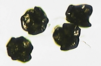
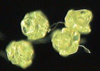
Certainly much of the pollen I collected direct from the flowers and examined dry were difficult to interpret and looked shrivelled. A good example is the American willowherb shown below (Epilobium ciliatum, an introduced and now widespread plant in the UK). The difference between the untreated and treated pollen is quite striking. (Although it is worth studying pollen from fresh anthers before treatment, especially to see the pollen colour).
It's worthwhile viewing the washed grains (still in a little alcohol) under a stereo microscope (ca 40X or greater for larger pollen types) as the 50% glycerine is added; the hydration and swelling of the grains is interesting to watch. Studying the hydrated larger pollen grains under the stereo also gives an idea of their gross shape and structure before studying at higher powers under the compound microscope.
|
|
|
|
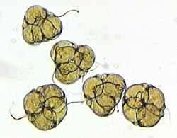
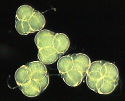
All pollen grains develop initially in groups of four called tetrads. In some plant species the grains separate; others don't. I haven't found a database image to check yet, but the tetrahedral form of the above willowherb pollen suggests this species is one where the tetrad doesn't separate. The willowherb species below has separate pollen grains.
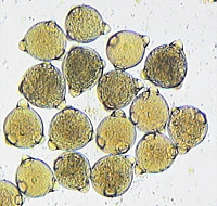
Rose-bay willowherb (Chamerion angustifolium). Brightfield (9X objective). Diameter ca. 80µm.
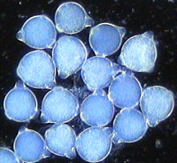
The same pollen under darkfield illumination. The pollen did look a blue green when collected.
Apparently flower pollen can vary in size from typically 3
micrometres (alpine forget-me-not, Myosotis alpestris)
to 200 microns (cucumber flower, Cucurbita pepo). Part
of the attraction of studying pollen is that the grains of
different species have a wide variety of shapes, structures and
surface ornamentation/pores. Pollen grains present a variety of
challenges to study and interpret under the microscope. Most
pollen requires at least a 10X if not up to a 40X objective and
higher to study both the gross structure and fine details. The
shallow depth of field at higher magnifications requires constant
refocussing and examining of grains in different orientations to
build up a mental picture of the pollen grain's shape and
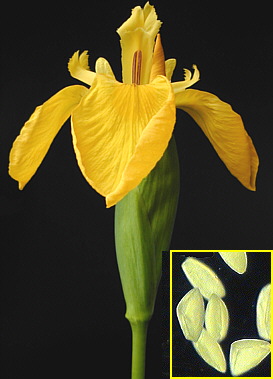
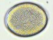
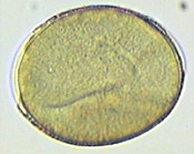
structure. The yellow iris pollen below demonstrates this.This
pollen was washed and hydrated and has swelled when compared with
the untreated iris pollen in the title image.The outer coat of a
pollen grain is called the exine and often shows fine structure.
|
|
The study of pollen and spores is part of the science of palynology, and is a well established field used in many branches of science and technology including forensic studies, vegetation studies (past and present) and oil exploration. Pollen and spores have a resistant coating and can be identified in the fossil record and from e.g. cores deep into peat bogs.
If the study of pollen appeals, it is worth doing a systematic study and perhaps build up a collection of permanent mounts e.g. of the common flowers local to you as they come into season. Unlike many botanical subjects where sectioning is required, making permanent mounts of pollen is fairly straightforward and there are some excellent guides and suppliers of materials to get you started (see references).
Identification of the pollen source is of course vital. There are many excellent garden flower and wild flower guides available nowadays to help with this. To ensure the pollen has come from the flower itself and not been deposited from elsewhere, pollen can be collected from buds that have been allowed to bloom and the anthers ripen away from contaminating sources (e.g. pine and grasses which can release copious amounts of pollen). It's worth noting the colour of the pollen when collected and removed from the anthers as further treatment and staining may well change this.
The preparation of the pollen for permanent mounts is minimal; after the treatment in alcohol and hydration mentioned above, one method is to then mount in glycerine jelly. NBS in the UK supply a special grade of glycerine jelly for mounting pollen which contains a red dye. This is adsorbed into the grains to show surface detail more clearly.
So if like the author you haven't studied pollen grains in any detail before, why not try studying some of the flowers in your garden or locally common wild flowers.
Comments to the author Dave Walker welcomed.
Image details. Pollen grains taken with Russian achromatic 9x and 20x objectives, homemade darkfield patch stops. Image capture without eyepiece with Panasonic CL-350 video camera and Snappy 2.0 capture card. Macro shot of lily with Fuji DX-10 digital camera.
References and further reading
1) 'Pollen, its collection and preparation for the microscope' by John White, UK. This privately published 37 page illustrated handbook is an excellent guide for the amateur naturalist/microscopist. Includes a list of reference books for further work. Available from Brunel Microscopes, UK.
2) 'Practical Microscopy' by Eric Marson, Northern Biological Supplies, UK. Booklet two in this bound set covers the preparation of mounts in glycerine jelly including pollen grains. The handbook covers a wide range of techniques for the amateur microscopist to prepare permanent mounts of many subjects. Available from Brunel Microscopes, UK.
3) On-line resources - a selection.
http://www.geo.arizona.edu/palynology/pol_pix.html 'Pictures of pollen grains on the web'. A useful compilation of links to pollen images on the web.
http://scrl.usda.gov/scrl/apmru/imms/pollen/reference_coll/Pollpics.htm An extensive collection of pollen grain images (both with the SEM and light microscope). Arranged by plant family. SEM images are particularly striking as they capture the depth of field and detail in one image that is difficult with light microscopes.
http://www.pollen.com For the unfortunate sufferers of hay fever, pollen is not a fun topic! This site covers the allergenic aspects of pollen as well as including images of particularly troublesome pollen and background on pollen counting techniques.
http://koning.ecsu.ctstateu.edu/plants_human/pollenemb.html Koning, Ross E. "Pollen and Embryo Sac". Plant Physiology Website. 1994. Part of an on-line biology course. Illustrates and describes the structure and function of the pollen grain.
http://ourworld.compuserve.com/homepages/Beekeeping/weblinks.htm This is an extensive listing of web resources for bee-keepers, including the many aspects of pollen that are of interest to them.
Published in the July 1999 edition of Micscape Magazine.
Please report any
Web problems or offer general comments to the Micscape Editor,
via the contact on current Micscape Index.
Micscape is the
on-line monthly magazine of the Microscopy UK web
site at Microscopy-UK