TRICHINOSCOPES.
by Mike Dingley - Australia.
There have been a few microscopes that have been designed and made for
specific tasks. One thinks of polarizing microscopes and dissecting microscopes
but how many of us know about the Trichinoscope?
Trichinella spiralis is a tiny nematode that is responsible for
the serious disease Trichinosis. Humans as well as dogs, cats, rats
and hogs can be infected and the cysts are found in poorly cooked pork
and dead rats. Nearly 75% of all rats are said to be infected.
LIFE CYCLE.
In pigs adult worms burrow into the mucosa of the small intestine where
the female produces larvae. The larvae penetrate into blood vessels and
are then carried, via the blood, to the skeletal muscles where they coil
up to form a characteristic-looking coiled cyst. This becomes calcified
and the adult worms can live here for years if undisturbed. When meat is
eaten the trichina enter the stomach and pass into the small intestine
and finally into the muscle tissue. Here they live and multiply in continuous
series, while the surrounding structures as well as the muscular tissue
undergo a process of histolysis. The destructive nature of the parasite
is very great. The number of progeny produced by one female may amount
to several thousands, and as soon as they leave the egg they either penetrate
through the blood vessels or are carried by the circulation until they
become lodged in the muscles.
Free Trichinella were seen for the first time by Zenker in 1860
and Rudolph Virchow (1821-1902) succeeded in inducing various German states
to make the testing of pig's meat for trichinosis in abattoirs compulsory
(c. 1870).
MICROSCOPES.
During my research into portable microscopes for a forthcoming book
I became aware of microscopes, or more formally called 'Trichinoscopes'
which are designed to look specifically for the trichines in meat. Although
I have not been able to find out very much information on them I thought
that readers might like to read an introduction to these little-known instruments.
There may be readers who might be able to pass on to me information on
the following instruments and others that I have not included here and
I will give full credit where due.
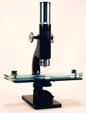
All of the instruments that I have seen have been monocular, low power,
compound and having characteristically large stages. The stages hold large
double glass plates between which is placed the meat to be examined. One
plate has a scale or grid etched into it and both plates can be squashed
together using screw nuts on either end of the plates. Compressing the
sample makes it thin and translucent.
The earliest instrument that I have managed to find is that depicted
in the Billings catalogue in Figure 146, made by Schmidt & Haensch,
Berlin in about 1879. It has a horseshoe base, rectangular pillar and stage
plate. The stage can be moved in a lateral motion as well as backwards
and forwards. Two glass plates 23.5cm X 4.1cm rest on the stage and the
lower plate is marked with five 2.54cm squares.
Figure 153, also in Billings, shows an instrument of unknown maker dated
before 1880. This instrument has a hardwood base 30.5cm X 10.5cm that also
acts as the stage. The two glass-plate compressoria measure 23.5cm X 4.1cm.
 Winkel-Zeiss
(1911-1935) made the Trichinen-Mikroskop as a travelling field microscope.
The author has the one in his collection. It is a black and chrome monocular
microscope with a large stage (20cm X 9cm). The single plane mirror is
on a swinging arm system. The eyepiece is marked T9X and it has coarse
focus back rack and pinion. The objective has a RMS thread (S/N 91504)
but the barrel is 30mm in diameter and has a lever which allows for two
magnifications; 4.5X and 11X. The two glass compressoria are 5mm thick
and 23cm long X 5cm wide. One has a grid etched on and are numbered 1-28.
The other piece is plain. The instrument is packed in an externally metal-lined
wooden case with lid and measures 30cm X 20.4cm X 9.7cm and has a leather
carry handle.
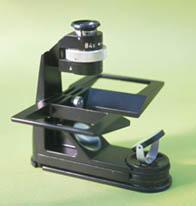
 PZO
the Polish Optical Company are still making (as at 1999) a small portable
trichinoscope called the Mtr. This is a splendid little instrument and
has an unusual focussing arrangement. The monocular eyepiece has a knurled
ring which, when rotated, gives a zoom magnification of X40-X84. The two
part stage folds up to surround the eyepiece and the base has two winged
feet which can be pulled out from beneath the base for extra support. The
glass plates measure 9.8cm X 4.5cm X .5cm and are etched with two layers
of grids marked 1-14. The microscope has a grey plastic top which fits
over the microscope and rests on a lip on the base to totally enclose the
instrument. The microscope is stored in a brown leather case which also
contains the many accessories that will be found useful in the field. The
glass plates rest on the two winged-stage which are then raised by hand
in order to focus the specimen. The dimensions of this little microscope
in its closed form are 3.8cm X 12cm X 12cm.
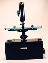
 H.
Hauptner, Berlin-Solingen. Black wooden box 11.7cm X 24.5cm
X 13cm with a carry handle. A boss is mounted on the lid. A key lock and
key is present under which is a label H. HAUPTNER Instrumentenfabrik BERLIN-SOLINGEN.
The end of the hinged pillar slots in to the boss in the case which also
acts as the base of the instrument. The single plane mirror is mounted
on a yoke.
The black painted-on-brass stage 17.5cm X 7.5cm slides on to a narrow
fixed stage plate with dovetail sides. Below this is a circular four hole
diaphragm. There are two glass specimen plates 23cm X 5cm X 0.7cm held
together with two nickel-plated screws. One plates is engraved with the
numbers 1-28 and has H. HAUPTNER, Berlin, NW7 marked. Rack and pinion coarse
focus is by a single knob. A single unmarked eyepiece and a non RMS thread
objective are present. The objective is unusual in that it has a lower
pivoting objective so as to give two magnifications. The case has a small
wooden box which contains three dissecting needles in wooden handles. Overall
height with the microscope on the case is 35.5cm and wight is about 3kg.
The rear of the pillar is stamped TM W1.
An instrument has been seen having the name KORTH
inscribed on it appears identical to the Hauptner instrument. There have
also been seen instruments very similar if not identical to the Hauptner
which are unsigned and therefore possibly manufactured by the same company
and distributed to various sellers. Without being able to personally inspect
these instruments it is very difficult to make judgments on their origins.
Another instrument made by MEOPTA in the 1950's
has come to light but at this stage the author has not been able to get
more information.
Comments to the author Mike
Dingley welcomed.
References:
Billings Microscope Collection of the Medical
Museum of the Armed Forces Institute of Pathology. The Armed Forces
Institue of Pathology, 2nd ed., 1974.
Hickman, C.P., Hickman, C.P. and Hickman, F.M.
Integrated Principles of Zoology. 5th Edition, 1974.
Hogg, J. The Microscope. Its History,
Construction, and Application. 15th edition, 1898.
© Microscopy UK or their contributors.
Published in the July 1999 edition of Micscape Magazine.
Please report any Web problems or offer general comments
to the Micscape
Editor,
via the contact on current Micscape Index.
Micscape is the on-line monthly magazine of the Microscopy
UK web
site at Microscopy-UK
WIDTH=1
© Onview.net Ltd, Microscopy-UK, and all contributors 1995 onwards. All rights
reserved. Main site is at www.microscopy-uk.org.uk with full mirror at www.microscopy-uk.net.
 Winkel-Zeiss
(1911-1935) made the Trichinen-Mikroskop as a travelling field microscope.
The author has the one in his collection. It is a black and chrome monocular
microscope with a large stage (20cm X 9cm). The single plane mirror is
on a swinging arm system. The eyepiece is marked T9X and it has coarse
focus back rack and pinion. The objective has a RMS thread (S/N 91504)
but the barrel is 30mm in diameter and has a lever which allows for two
magnifications; 4.5X and 11X. The two glass compressoria are 5mm thick
and 23cm long X 5cm wide. One has a grid etched on and are numbered 1-28.
The other piece is plain. The instrument is packed in an externally metal-lined
wooden case with lid and measures 30cm X 20.4cm X 9.7cm and has a leather
carry handle.
 PZO
the Polish Optical Company are still making (as at 1999) a small portable
trichinoscope called the Mtr. This is a splendid little instrument and
has an unusual focussing arrangement. The monocular eyepiece has a knurled
ring which, when rotated, gives a zoom magnification of X40-X84. The two
part stage folds up to surround the eyepiece and the base has two winged
feet which can be pulled out from beneath the base for extra support. The
glass plates measure 9.8cm X 4.5cm X .5cm and are etched with two layers
of grids marked 1-14. The microscope has a grey plastic top which fits
over the microscope and rests on a lip on the base to totally enclose the
instrument. The microscope is stored in a brown leather case which also
contains the many accessories that will be found useful in the field. The
glass plates rest on the two winged-stage which are then raised by hand
in order to focus the specimen. The dimensions of this little microscope
in its closed form are 3.8cm X 12cm X 12cm.
 H.
Hauptner, Berlin-Solingen. Black wooden box 11.7cm X 24.5cm
X 13cm with a carry handle. A boss is mounted on the lid. A key lock and
key is present under which is a label H. HAUPTNER Instrumentenfabrik BERLIN-SOLINGEN.
The end of the hinged pillar slots in to the boss in the case which also
acts as the base of the instrument. The single plane mirror is mounted
on a yoke.