Hang on, you’re in for a bit of pun-ishment. As I concluded in an earlier essay, even when we are agitated Nature provides us Psolus.
Or when we take that radical metaphysical view that others really don’t exist, this is the sea cucumber position known as Psolupsism or when we commit a major verbal gaff, then we are guilty of a Psolecism. Sorry about that, I will now go off and do penance in Psolitude. My wife and friends agree and have strongly demonstrated their Psoludarity.
O.K., let’s start over and put all of that behind us, because Psolus really is a fascinating organism. Although many echinoderms demonstrate a pentamorous or five-fold symmetry–think of the “ordinary” starfish–it is certainly not obvious in holothuroids (sea cucumbers). However, in a considerable number of species, careful observation will reveal such a symmetry–especially internally. Biologists sometimes have an odd way of looking at things and a peculiar way of counting, but once you understand what’s going on in the organisms, you begin to understand why. (Maybe.) In lots of species, if you look at the bundles of tentacles (or as some call them “buccal podia”–in other words, they are special modified feet around the mouth), you will find 10 or as the taxonomic morphologist will tell us, 5 sets of pairs. When you investigate the internal anatomy, you will find that such a model is justified. In sea cucumbers, we ordinarily find the tentacles at the “front” end and the anus at the “rear” end. (In some species, there are anal “teeth”–I’ll bet Freud would have had fun with that.)
The preserved specimens which I possess, have, of course, contracted and lost much of their color. In live specimens, the tentacles are a bright red and can present a colorful display. However, no beauty contest winners here especially among the preserved specimens. Here’s a link that will let you see what they look like when alive.
Now, my specimens are not as attractive, but they are interesting.
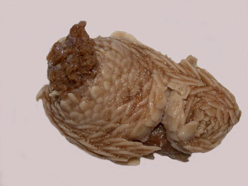
Here, as you can see, the tentacles remain partially extended and the plates or scales on the body are clearly visible.
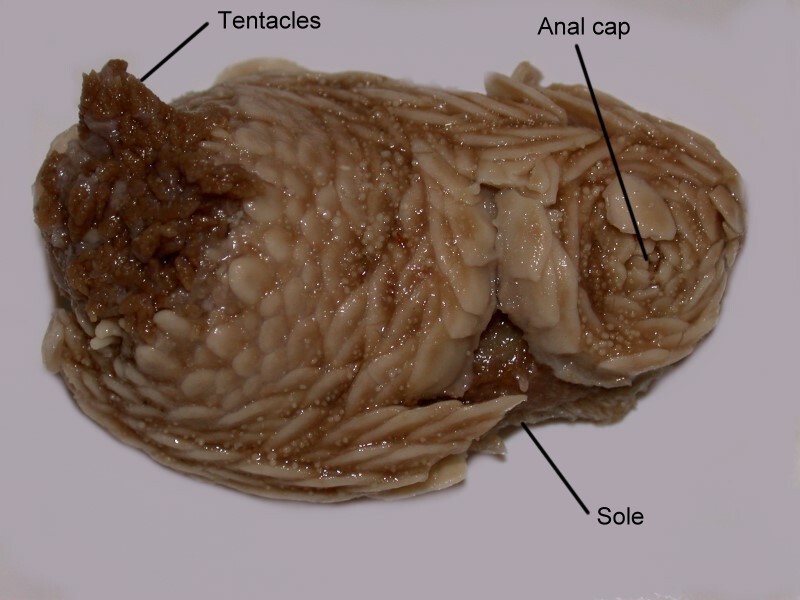
In this image, you can see, not only the tentacles, but the set of plates which form the “anal cap.”
The armor of this creatures is really quite remarkable as we can see in this view of its posterior.
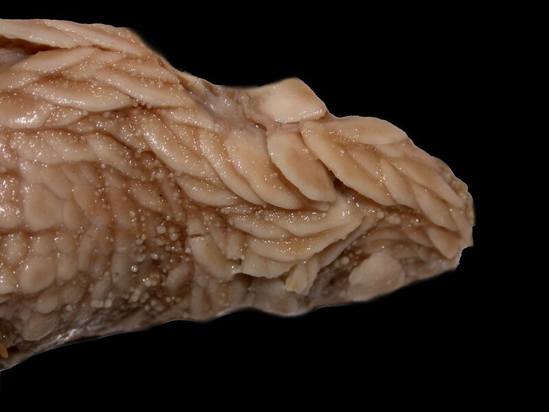
There is a heavy layering of connected scales forming what I am going to refer to as a “collar” around the tentacles.
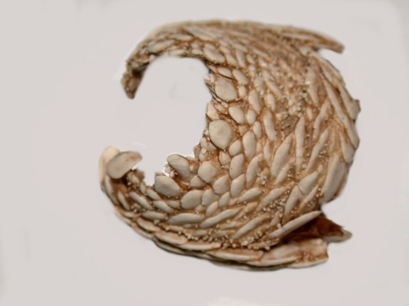
When I began to dissect, I removed this collar intact and also the “anal cap” which looks rather like a stone flower as you can see below.
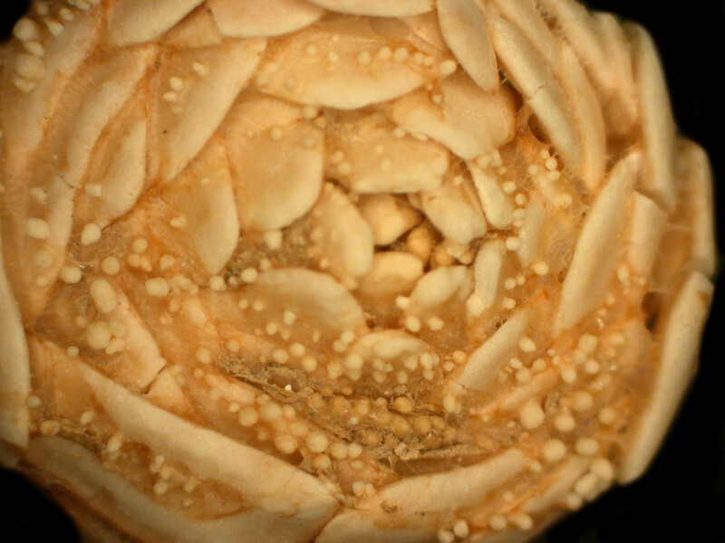
You will notice that, in addition to the plates or scales, there are many small spherules. These structures are all calcareous, but some are more clearly crystalline in composition than others.
When we look at the underside of the anal cap, we see a Maltese cross. This is something of a surprise , since echinoderm symmetry is most commonly pentamorous rather than quadrate.
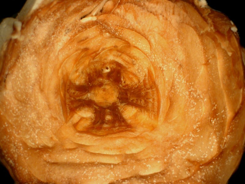
This anal cap may seem a bit of an overreaction, but some sea cucumbers have reason to be a bit nervous about invasion of their private parts. There is a small fish, popularly known as a pearl fish, at least 2 genera of which are known to enter the anus of certain species of sea cucumbers and take shelter in the respiratory trees.
Even though these creatures are usually sausage-shaped, there is generally a dorsal and ventral aspect. The ordinary pattern is that of 3 sets of pairs of tube feet on the dorsal (top or “back” side). What’s particularly interesting here is that the 3 ventral sets (the “trivium”) are locomotory and tend to use suction, whereas the 2 dorsal sets (the “bivium”) have podia which are papillate and are fundamentally sensory in character.
In Psolus, the ventral side is flattened into two leathery “soles” with a thin strip of tissue dividing them so that the right side looks like an elongate “D” and the left side is its mirror image.
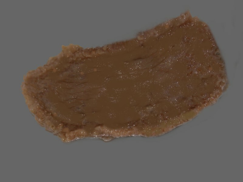
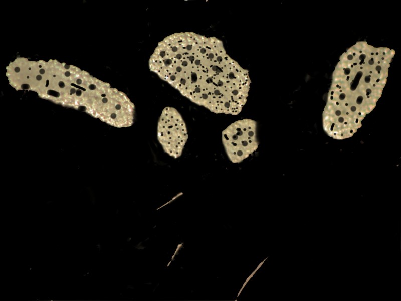
There is also a strip around the entire perimeter of the ventral side which has a series of small pouches and also tube feet for locomotion. These tube feet do have ossicles and they are highly fenestrated.
This ventral sole, in some species, serves as a sort of nursery in which embryonic forms develop and are protected. [There is a drawing in Libbie Hyman’s volume IV (Echinodermata) of the 6 volume set The Invertebrates, page 187, Fig. 78A. ] This remarkable drawing (after Ludwig) shows 22 larval forms on the underside. In the drawing, where the species is Psolus antarcticus, you can see the tiny sea cucumbers already protected by an armor of scales. As this splendid plate shows, at least 2 other species of Psolus and a species of Cucumaria have brood pockets. In Psolus koehleri, there are 5 brood pockets around the neck and, in Cucumaria crocea, 2 pockets in the neck, and in Psolus ephippifer, there are brood pockets covered by calcareous plates.
I have 6 specimens of the species Psolus fabricii which were collected in Maine. I have not yet confirmed that this species has brood pockets; if I can determine whether or not it does, I’ll let you know. At the bottom of the jar is a considerable amount of debris, largely material cast off by these organisms. Some species of sea cucumbers are notorious for eviscerating themselves when they are disturbed or attacked, often leaving the attacker entangled in a sticky network of viscera. If, subsequently, the sea cucumbers are able to find favorable conditions, they will regenerate the parts which they have ejected.
In this debris, I found a number of the types of “scales” which almost entirely cover the dorsal aspect of Psolus and are also found around the margin of the ventral side as well. These calcareous plates have a fairly complex arrangement and there is overlapping, so that this creature is rather well armored, as we have seen.
I was examining one of these plates from the debris and the underside which was attached to the body, is flat, clearly crystalline in nature and rhomboidal in shape. I expected that when I turned it over I would find basically the same thing–surprise! The outer surface was covered with tissue–well, that’s no surprise–and the appearance was that of tiny warts in the tissue–well, that’s no surprise either; just take a good took at Stichopus. The surprise is that these warts are not composed of tissue. I was suspicious because the light patterns suggested a distinct difference between the surrounding tissue and the warts. Using some of my wonderful micro-tools–I keep telling my friends “You can never have too many micro-tools!”–I scraped off some of the tissue with its warts and subjected it to a brief treatment in Clorox. (In order to minimize the time of exposure to the bleach, I now dissect away as much extraneous tissue as possible and then, while the item of interest is in the bleach, use micro-needles to accelerate the removal of the tissue, so that as quickly as possible, I can transfer my specimens to distilled water rinses.) Now, here’s the surprise; these warts turn out to be small, roundly spherical calcareous structures. I’m sure somebody has noted, described, and named these structures before, but, so far, I have been unable to find any references to them.
The next step was to place a couple of scales which were still covered with some tissue containing some of the spherules in Clorox. I put the material in a small Petri dish with enough fluid to thoroughly cover the specimens and a bit more so that I wouldn’t confront excessive evaporation that would leave my scales embedded in a crystalline lump of sodium hypochlorite. I let them sit overnight and when I went to examine them the next afternoon, I got 2 more surprises. Not quite all the tissue had dissolved away and this turned out to be a distinct advantage. This is a little tricky, so hang in there and I’ll try to make this as clear as possible.
First of all, there were some small sheets of tissue which no longer had any calcareous material, but looked as though someone had taken a micro-paper punch and made a series of small, round holes in it.
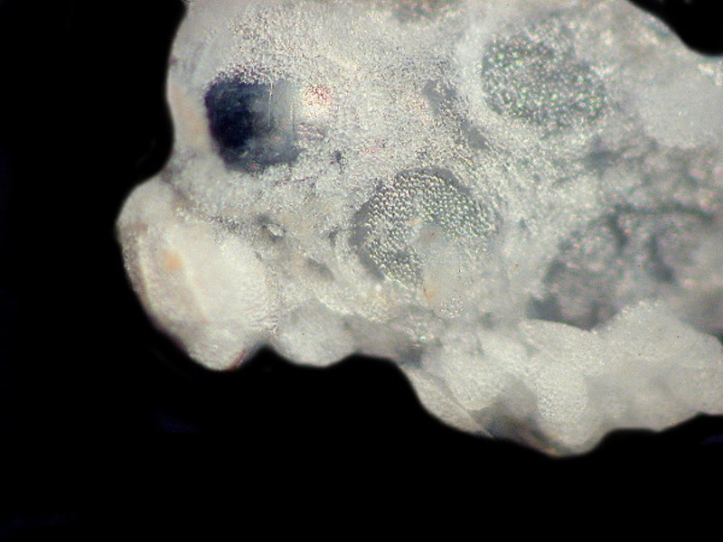
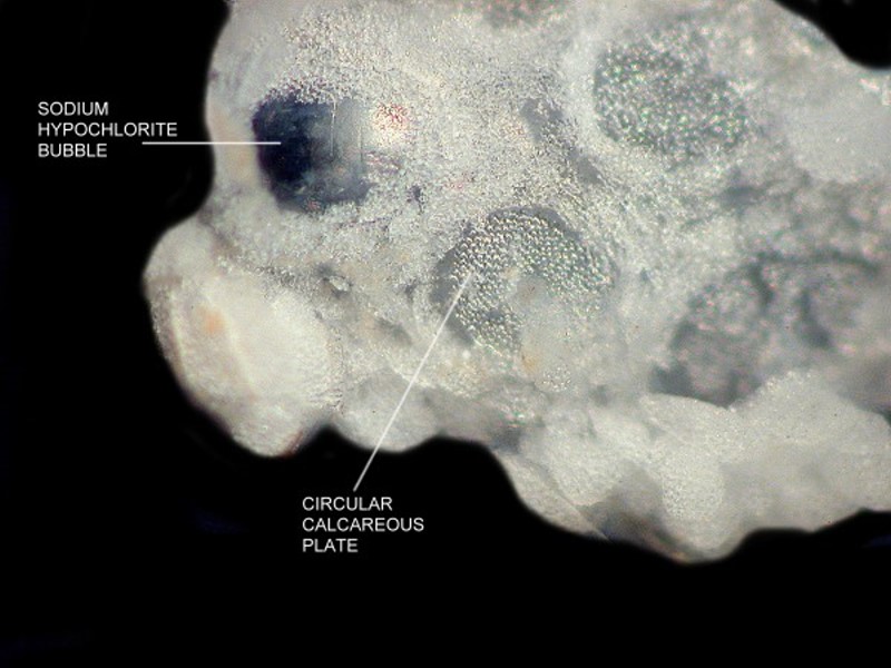
Secondly, there were a couple of pieces of tissue that still had some calcareous material. I assumed that here I would find spherules–there were plenty of them around in the bottom of the dish–but, instead, what I found was a flat, round disk composed of a series of small fenestrated plates. It is at this point that partial retention of bits of the surrounding tissue gets really interesting and valuable. Surrounding this circular disk is a series of very small, flat fenestrated plates which are almost like a miniature fence around the central disk–curiouser and curiouser! When I used my micro-needles to dissect away bits of the tissue, the entire edifice began falling away into its component parts. So where do the spherules fit in? I still don’t yet know.
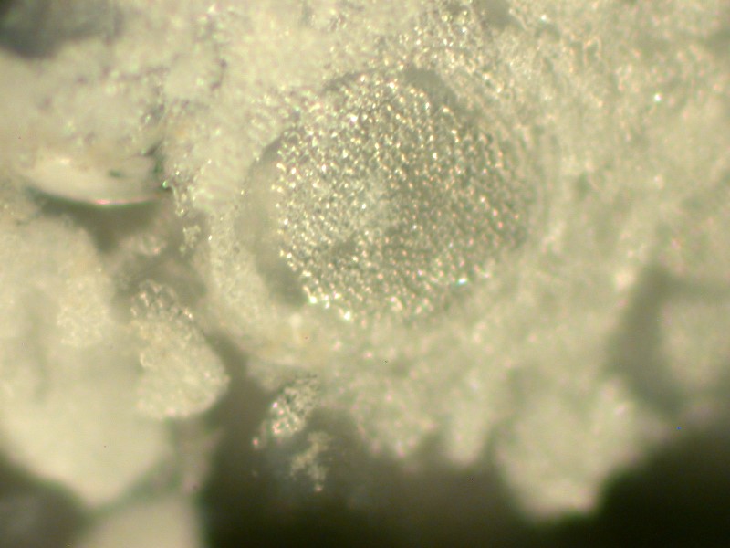
Thirdly, I began to examine one of the scales with a sense of confidence that here I would find a nice, neat stable rhomboidal crystalline entity–surprise! As soon as the edge of the scale came into view, I knew that new mysteries were afoot.
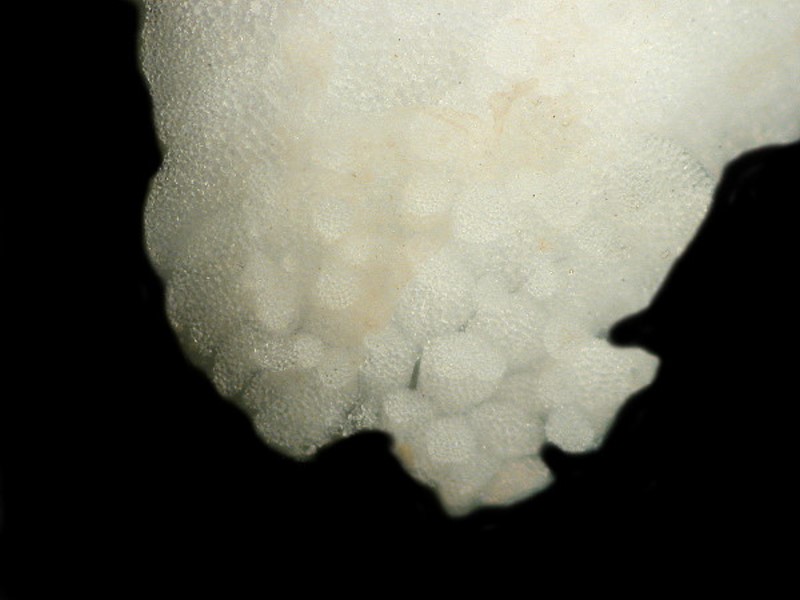
There were several spherules around the edge and as soon as I touched the scale with the micro-needle, fenestrated plates in a variety of shapes and sizes, spherules, and thin flat plates began to fall off. Hundreds of pieces in each scale! So, how does the architecture here work? Well, it turns out that I didn’t get that quite right, since I went back and poked around some more and found that some scales are quite stable rhomboids and are not composed of other little bits and pieces which simply adds a new puzzling dimension.
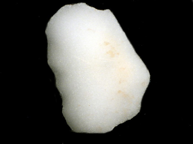
At this point (but it’s still early days), it seems to me that this creature seems to “stack and pack” in building its armor. Something I find rather curious is the production of all these different sizes and shapes; it’s a bit like a miniature calcareous tile factory. The technical term for this kind of overlapping is “imbrication” but, surely something more is going on than mere overlapping with all these size variations and the areas that are densely packed either with fenestrated platelets or spherules. I’ll continue looking.
Another oddity about Psolus is that the tentacles (or buccal podia) are not at the anterior end, but arise out of a dorsal introvert which it can extend and retract and furthermore the tentacles are capable of exuding a toxic substance which tends to keep other organisms from nibbling on them. In fact, almost all sea cucumbers secrete a toxic slime to discourage predators and, in general, it works quite well, the major exception being humans who collect several thousand tons of sea cucumbers annually as food. The organisms are slit open and often boiled to remove the salt and toxins and then they are dried. This material is generally described by the Malay word ‘trepang’. It is often cut into cubes and used in soups and stews. Have I tried it? You’ve got to be kidding; I have a very conservative 77 year old stomach! I think it may be not only an acquired taste, but a genetic one, since the dominant consumers are either Asian Polynesian, or Eskimo. Trepang has not succeeded in becoming a Western food fad.
When I begin to examine a complete Psolus bit by bit, I will also be looking for commensals and parasites, since most organisms have these and even some parasites have parasites. Yesterday, I happened across a terse account stating that the dorsal commensals of Psolus tend to have the coloring and general appearance of the scales. I find such remarks intriguing and exasperating, for while they are provocative, the fact that no examples or evidence is offered can lead one off on a wild goose chase. It also happens to be true that many parasites and commensals are adept at camouflage which simply makes the whole issue even more intriguing and more exasperating.
This afternoon, I was again looking at some scale detritus and I noticed on some tissue surrounding the plates and spherules several items of interest. The first was the lightly blue-tinted lorica (tube house) of a protozoan known as Folliculina. Back in my pre-digital Polaroid days, I found some loricas of this organism on the tunicate Styela plicata with the contracted organisms still in them. Here’s a view.
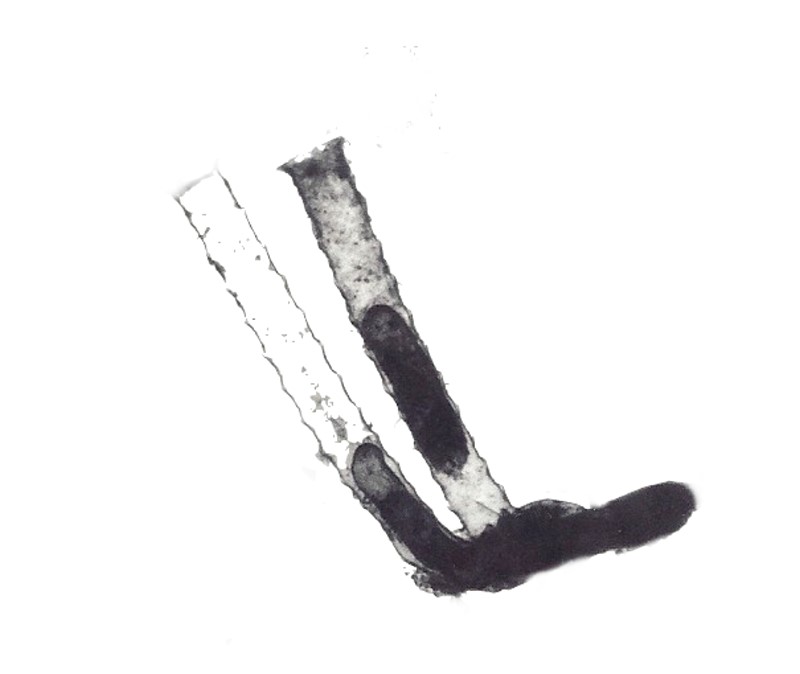
This is an elegant, interesting creature, so if you’re fortunate enough to live near an ocean or sea, do look for it. It’s fairly common and quite unmistakable. The second thing I noticed was a number of the highly fenestrated plates embedded at various angles in the tissue. There didn’t seem to be any particular order, but that may well have simply been a consequence of the general disarray of the material in the detritus. The third, and to me, most interesting item was a series of transparent rods in the tissue; some were short, some of relatively medium size, some slightly curved and pointed at the ends. They very much remind me of certain monaxon sponge spicules but, I am quite sure that these actually belong to the Psolus, so I'll probably be wrong and if I am I’ll let you know. A few of them appeared to have what might be a slightly rounded terminus at one end, but I won’t be able to determine that until I examine them under a compound microscope.
I decided that I should isolate a few of the rods and subject them to the bleach test and the acid test. These are 2 basic, simple tests, but they are often very helpful. The bleach test tends to rule out a wide variety of organic compounds. If, however, we were dealing with cellulosic compounds in tunicates, this test wouldn’t be much help, but if clear rods such as these we are examining dissolve in bleach, then that would be rather a surprise; as a matter of fact, they didn’t and remained quite stable. Then I put a few more rods on another slide and added a drop of 10% hydrochloric acid. (You never want to mix this with bleach unless you're willing to risk very serious chlorine poisoning!) If the rods start bubbling away and quietly dissolve, then they are calcareous; if they don’t there’s a very good chance that they are siliceous. These rods didn’t bubble, squeak, or dissolve, so I strongly suspect that they are silica which adds another puzzle to our series. In my years of being awed by echinoderms, I have never come across any siliceous structures nor have I read any account of such. So again, I am brought to wondering about rogue sponges that might be commensals of Psolus; that’s the simplest and most probable explanation but, since Mother Nature is such a prankster, I am reluctant to jump to any conclusions.
Feeding in Psolus poses another small puzzle since the tentacles are located on the dorsal introvert and they are short enough that it seems highly unlikely that they are extended down from the introvert over the body and onto the substrate to feed. Typically, sea cucumbers feed on almost anything in their path that is small enough to ingest which means that usually a large amount of sand and detritus passes through their gut. A few species are highly selective; for example there is one form that feeds almost exclusively on a particular kind of kelp but such connoisseurs are rare among holothuroids. Some species bury themselves in the substrate with just the tentacles protruding above the surface to feed on surround detritus and the “rain” of debris that is constantly falling through the water to settle on the bottom. Yet other species are burrowers and churn their way through the sand leaving trails. I suspect that Psolus is an intermittent burrower; the pattern of imbrication of the scales suggests that it could move fairly readily through a sandy substrate, find a suitable spot, extend its introvert, test the water with its tentacles and then feed or move on.
However, now I need to backtrack a bit. This afternoon, I isolated a tube foot and subjected it to bleach to see if it contained support structures, since not all echinoderm podia do. For example, I also placed a tube from a small starfish in bleach and it dissolved completely–no skeletal structures in that part. Some sea urchins have a beautiful arrangement of calcareous plates which form a disk in the tube foot and you can see an example below from Strongylocentrotus franciscanus.
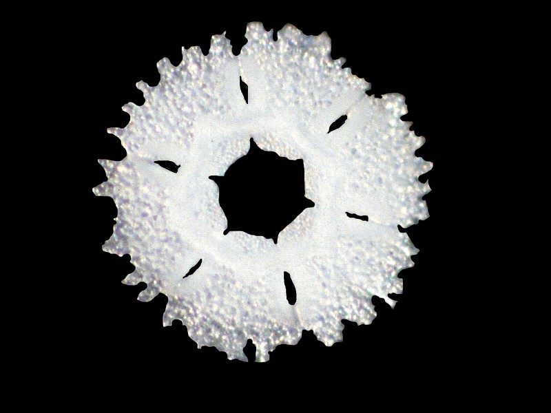
The disk is composed of six plates which are delicately joined together. Below you is an example of two of the plates which comprise the disk.
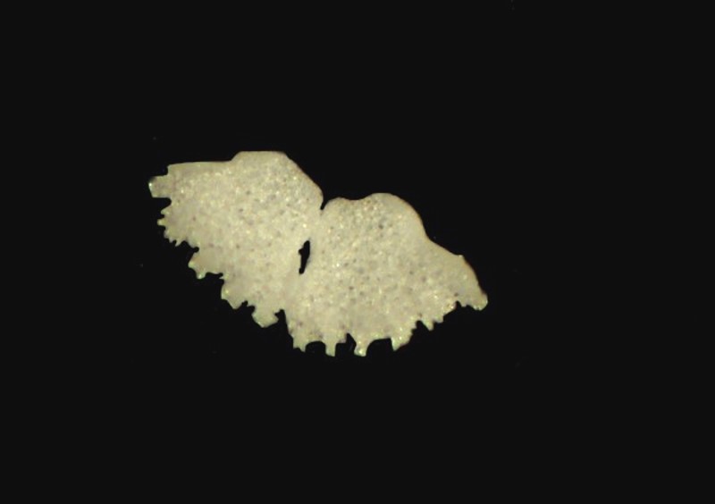
Furthermore, just under the disk, there is an hexagonal support form which you can see here in isolation
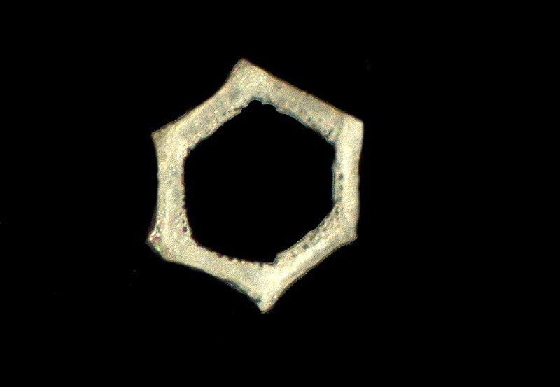
and again incorporated with the disk.
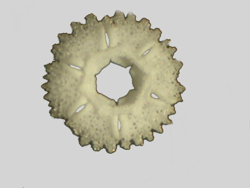
Psolus fabricii does not have any such elegant podial arrangements, but it does have fenestrated plates and rods.
Here the plates are scattered and it may be that ordinarily they have a more coherent arrangement, but that remains to be determined. The calcareous plates were not a surprise, but the presence of siliceous rods was, so some cautious rethinking is necessary.
As is often the case, I have rambled on making this essay already rather long. I will follow up with another article expanding some of my observations on Psolus along with some investigations of some other small sea cucumbers from the Philippines.
All comments to the author Richard Howey are welcomed.
Editor's note: Visit Richard Howey's new website at http://rhowey.googlepages.com/home where he plans to share aspects of his wide interests.
Microscopy UK Front
Page
Micscape
Magazine
Article
Library
© Microscopy UK or their contributors.
Published in the July 2015 edition of Micscape Magazine.
Please report any Web problems or offer general comments to the Micscape Editor .
Micscape is the on-line monthly magazine of the Microscopy UK website at Microscopy-UK .
©
Onview.net Ltd, Microscopy-UK, and all contributors 1995
onwards. All rights reserved.
Main site is at
www.microscopy-uk.org.uk .