| Vacuoles, Form and
Cannibalism? A collection of
miscellaneous images
By Paul James, UK
|
Clearly it seems
that one of the very first hurdles encountered by the emerging
unicellular life forms was the problem of dumping excess water
that seeped into the cell by the osmotic process. The established
method : that of collecting this unwanted water into an expanding
'vacuole' ready for pumping back out evolved to deal with this
rapid hydration. The novice soon learns to spot the presence of
these ubiquitous cavities quite quickly throughout the huge range
of protists that soil/pond water hosts.
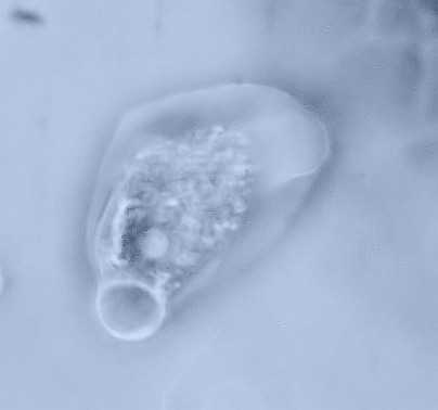
Sometimes it is not
easy to distinguish between water and food vacuoles, but all
unicellular life forms must have to rid themselves of excess
water, so if only one vacuole is present at the moment of
observation it is likely to be one dealing with the osmotic
problem. Food vacuoles are not always present of course, and the
various species of amoeba deal with food particles in slightly
different ways. Often we see particles of food tumbling inside
beside the rest of the cell contents without any apparent
self-containment inside a vacuole. Some varieties have more than
one water vacuole which often fuse together into a very large
cavity before expelling the water through the ectoplasm, whilst
others appear to constantly possess several simultaneously.
I thought of
posting a few images of amoeba gleaned over the years which
illustrate the variation water vacuoles take in size, position
and number. The soil/pond water enthusiast soon learns that there
are many amoeboid varieties throughout the species, ( literally
thousands ) a few of which become familiar to the eye over the
years. My own home patch sports various forms which I give simple
personalised names such as the 'Carpet Slipper' ...... '
Balloon'......'Skull'......' Formula 1' etc. as I recognise them
during observation !
 |
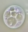
Water or food vacuole
?.......... it's large for a tiny amoeba
|
 |
| Two water vacuoles fusing into
one. Note the nucleus pinched between the
ectoplasm and vacuole.
|
 |
Almost perfect clock dial
formed symmetry from resting amoeba which nevertheless
has to continue pumping out
water regardless.
|
 |
| Curious form showing 'tail'
water vacuole imitating the neck of
a balloon.
|
 |
| Three large vacuoles in an
unusually rectangular cell form I've not seen before. |
 |
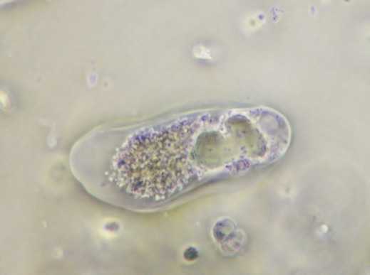
| Difficult to distinguish
between water and food vacuoles from a single image
like this. Prolonged observation will provide the answer
but can
take longer than expected for the water to
eject in some examples.
|
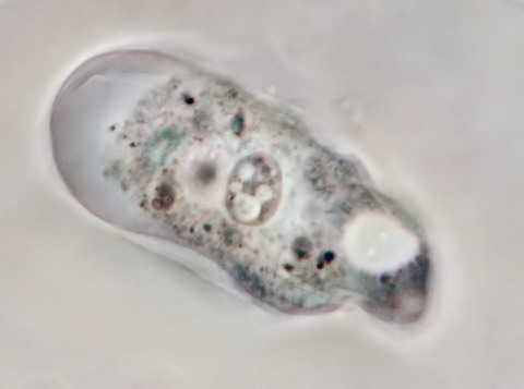
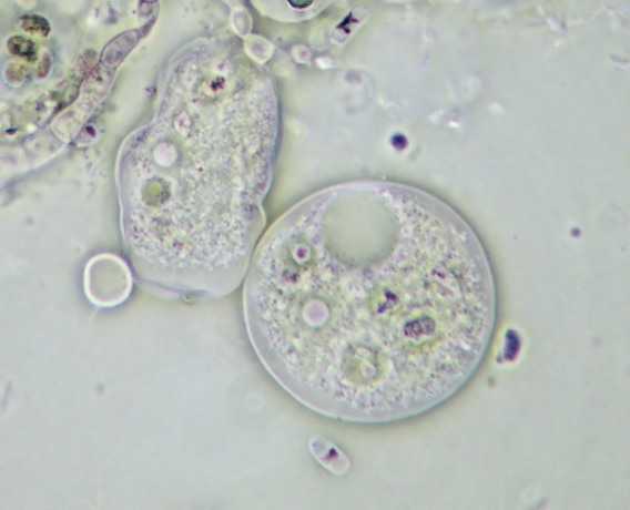
At around the time of collecting
together the images for this article during a session of
observation, I chanced up a rather curious sight of an a soil
amoeba which seemed to have engulfed another, albeit a lot
smaller. The best image of this from a large sequence recorded
onto my digicam's memory card is shown above, which incidentally
has been contrast enhanced to make the illustration of the
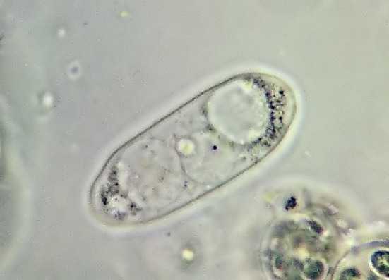
endoplasm a little clearer. On the right is an example of a tiny
soil amoeba at about the same amplification as the main image.
Though there is no doubt that there is a similarity between the
two, I cannot be certain that this is an example of 'cannabalism', but the ingested amoeboid form appears to be genuine. I
wonder how many other observers have noticed a similar feeding
habit ?
If my
images appear a little soft it is simply because I have left them
so and importantly removed all noise from them, then compressed
the files into small sub 10k packets. As a frustrated dial up
subscriber I have to consider my own uploading times, and no
doubt too hopefully at least, someone somewhere with similarly
poor web access might be glad of an article that's easily down
loaded.
Microscopy UK Front Page
Micscape
Magazine
Article
Library
© Microscopy UK or their
contributors.
Published in the July 2008 edition
of Micscape.
Please report any Web problems or
offer general comments to the
Micscape Editor.
Micscape is the on-line monthly
magazine of the Microscopy UK web
site at
Microscopy-UK