|
Slide query: Amphipleura
pellucida diatom slide dated 1950 by David Walker, UK |
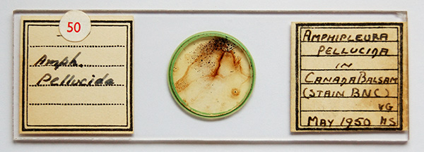
In my motley collection of diatom slides, one that intrigues me is a strew of the well known test diatom Amphipleura pellucida with a label stating that it has been stained with 'BNC'. The mounter is not stated but is dated May 1950 and used Canada Balsam mount. My own diatom studies are casual but was interested to know if BNC staining of diatom frustules is a known protocol or whether the mounter was just experimenting. In either case, any information on the nature of the stain 'BNC' would be welcomed as I can't find a reference to it.
In a wider context, this slide made me wonder as a hobbyist whether staining of diatom slides was a well established technique for optical microscopy studies as don't recall reading about it in the literature I have ready access to. High refractive index mountants have long being used to improve the contrast including the use of exotic mountants such as Realgar. Cleaned diatom frustules are primarily silica with the surface activity possibly dependent on how the frustules have been processed, so uncertain how well conventional stains would 'take' (for want of a more scientific description) to the diatoms. As the examples below show, the apparent 'take-up' of 'BNC' stain was extensive for many diatoms although very variable in this slide.
An image of the slide and photos of the strew with some observations are below. Any insight on the slide or of staining diatoms would be of interest. Thank you.

No
mounter is stated. The stain has left residual uneven streaking
and top right there are rectangular blocks of brown material, see below.
Although
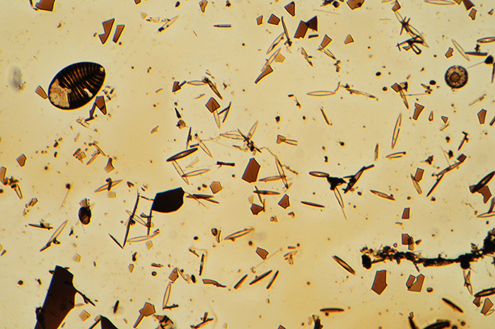
labelled as a strew of A. pellucida, a number of other species are also
present.

The
slide seen with a 6.3x objective. It's not an attractive slide! Irregular dark
brown fragments (stain and/or residue?) are present.

Objective 40x. Stain 'take up' was very variable between species and between examples of a species. Uneven staining within many specimens was also seen. The presence of staining outside a diatom border was commonly seen for denser stained examples. It's uncertain if this occurred either at the time of staining and/or leaching of stain into the mount afterwards.

When the staining level was at an intermediate density and even, to my eye the effect was quite pleasing, as in the above example.

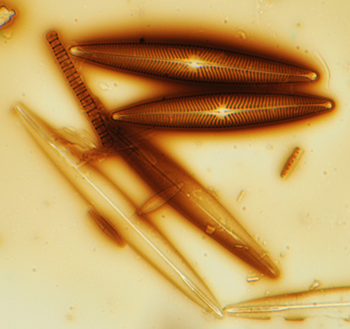
Objective 40x. The extensive staining outside the peripheries of some diatoms, as above, gave a rather surreal look to them.

Objective 100x, oblique. A close-up of the above species, but not the same example.

A.
pellucida, 100x objective, strong oblique along long axis.
Presumably
the original staining was to aid the resolution of the
fine detail on this classic test diatom, either striae and/or punctae.
But I struggled to get crisp images of fine detail of coarser
detailed species on the slide, possibly because of the thicker than
usual slide causing some aberration, which didn't bode well for
critical A. pellucida studies.
Striae were seen with oblique
on more lightly stained examples of A. pellucida as shown above.
Although I did try to compare the detail on stained specimens
with those of unstained examples on the slide, I found it hard to judge
whether the staining had the potential to aid the resolution of
fine detail.
Using blue light gave overly strong contrast and
evidence that the stain was nearly opaque to the shorter wavelengths,
as the stain fragments were near black in blue light.
So, overall, the slide seems rather an oddity to my casual eye, but an interesting one as had never previously come across a stained diatom slide.
Published in the July 2008 edition of Micscape.
Please report any Web problems or offer general comments to the Micscape Editor .
Micscape is the on-line monthly magazine of the Microscopy UK web site at Microscopy-UK
© Onview.net Ltd,
Microscopy-UK, and all contributors 1995 onwards. All rights reserved.
Main site is at www.microscopy-uk.org.uk with full
mirror at www.microscopy-uk.net .