|
THE
COCKROACHES
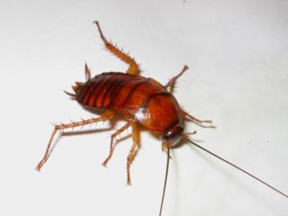
Cockroaches
are
endorsed by 350 million
years of life on earth. They are real "living fossils", as an enormous
amount of
species have existed since the Carboniferous period with a persistence
that
demonstrates their
successful and adaptable structure.
The
diversity and extent to which they are represented in that geological
era
clearly indicates that there was a long previous evolution of roaches
that has
left no vestiges. There are now more or less 3500 living wild species.
Nevertheless,
here we are interested mostly in those few species that have adapted to
the world
of the civilized man and to the human environment, becoming a plague
detested
by all housewives and owners of bars, restaurants, supermarkets and
food
depositories of all types; thanks
to their
fertility, adaptability to all kind of foods, and especially to their
cryptic
behavior that assures them of relatively few encounters with man.
|
|
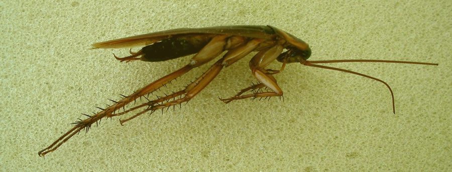
This
picture clearly show why the cockroaches are capable of colonizing very
narrow
cracks in floors, walls and furniture.
|
They adapt
easily
to most of the chemical pesticides that where intended to control them,
and
they end up developing resistance to them. So the modern way for the
safe
control
of cockroaches is turning out to be the old methods of physical control
and
the traditional
papers covered with sticky substances.
See
the Appendix for some old but
effective
control methods.
Throughout
the world the species of cockroaches that have been successful in man's
environment
are Periplaneta americana, Blatta orientalis, Blatella germanica,
Blaptica dubia, Suppella supellectillium, Suppella longipalpa, etc.
Except for Blaptica
dubia, from South
America, the other four
genera are perfectly adapted to Europe.
In Durango (Dgo.) Periplaneta
americana is common in the city,
living mainly in the culverts. We suffered in Hermosillo (Sonora) the invasion of the house by Blatella
germanica, and in Cancún (Quintana Roo) we have found up
until
now P. americana and Blatta orientalis.
You
do not
have to be deceived by the name of Periplaneta
americana. According to one author writing on the
internet, the origin and dispersion center of this highly invasive
species is Africa from where it arrived at the United States in 1625. Linnaeus, created in 1758
the genus Blatta in which he included
the species americana (surely because the available specimens were from America), and Burmesteir in 1838 created
the genus Periplaneta to hold americana.
HTTP://creatures.ifas.ufl.edu/urban/roaches/american_cockroach.htm
Readers
who
wish
to see images of the different species, can make use of their favorite
"search engine"
including in the search the complete species name, written between
quotes. Most
of the web literature is related to the “pest” control, and companies
offering their
services.
|
|
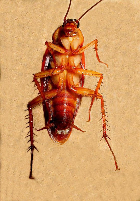
Male,
periplaneta,
ventral view, approx. 3x natural size.
|
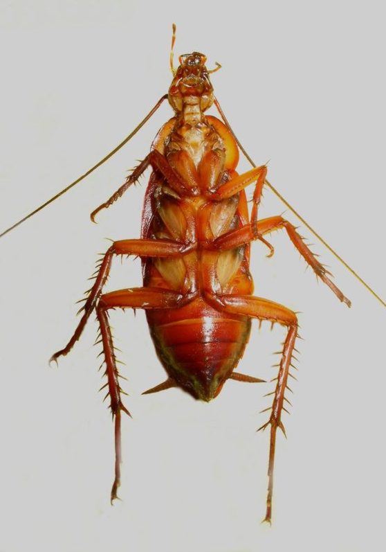
Female,
periplaneta,
ventral view, approx. 3x natural size.
|
Torrential rains
of this season (June-July) force the roaches to leave their hiding
places
even in
the daylight, terrifying the housewives that take care of their house
hygiene.
The
municipality
collaborates in the control of the plague by fumigating with
insecticide, with
very
little adoption of ecological criteria and techniques. This provided me
with
several specimens
to illustrate this article. If I wanted I could have had dozens at my
disposition.
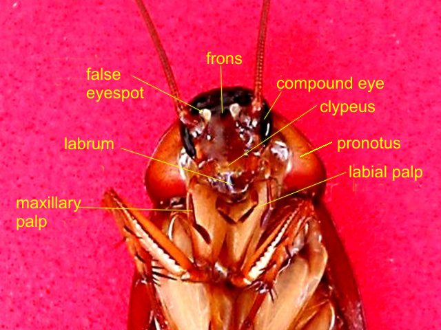
|
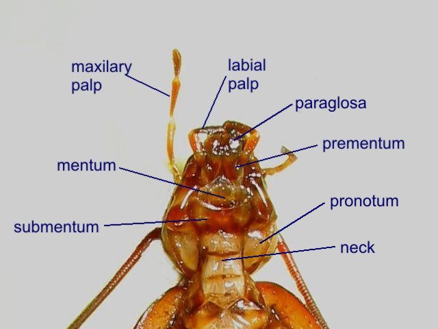
|
Periplaneta,
head, frontal view
|
Periplaneta,
head, ventral view
|
Whoever wants a more thorough in depth view on the world
of cockroaches, including those species that
some use as pets, like the sizzling cockroach of Madagascar, and other
but
smaller
species that herpetologists raise as food for their other pets
(lizards and
snakes), can visit these Internet addresses:
HTTP://www.uky.edu/Agriculture/Entomology/entfacts/misc/ef014.htm
HTTP://www.easyinsects.Co.uk/cockroaches/madagascan-hissing/index.HTML
HTTP://www.accessexcellence.org/RC/CT/roach.html
The anatomy
of a cockroach is a magnificent example of the structure of an insect.
This
explains why several educational pages on the internet describe it in
detail. The
best one by its didactic quality and beautiful macro and
photomicrographic
images, is this site of the University of Alberta:
HTTP://www.biology.ualberta.ca/facilities/multimedia/uploads/entomology/entposters.pdf
Also
a very
complete and descriptive site, but illustrated only with drawings, is
the one of
Prof. Fox, at the Lander
University site:
HTTP://www.to
lander.edu/rsfox/310PeriplanetaLab.HTML
The notes that
follow will serve as a guide for those who wish to try the
parasitologic
investigation of a cockroach.
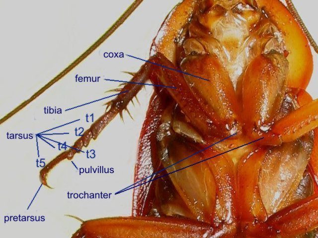
|
The cockroach leg is not
totally typical. Normally the coxa is a small segment and the
trochanter an important one; a design that is inversed in this case.
|
Objections
to dissection and justification of its use
If somebody
has ethical doubts on this type of operation he can find an important
discussion
of this subject at:
HTTP://www.elements.buap.mx/num46/htm/19.htm
HTTP://www.cahiers-antispecistes.org/article.php3?id_article=229
Many
very
religious people and strict vegetarians by, for example imitating San Benito, walk with care to ensure they don't squash
even an ant. But most of the non-religious or ideological objections
have resulted
in the avoidance of dissections in schools and colleges as they are
considered cruel and in bad taste, (which according to my own
experience at
high
school when 13 years old, is really sound). To avoid where
possible the extent of animal suffering under experimentation, when
that is inevitable
in scientific
research, suitable methods of anesthesia and euthanasia are used.
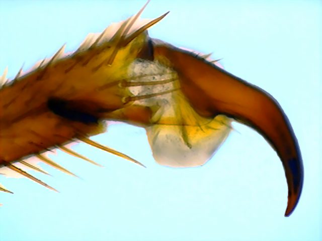
|
|
|
|
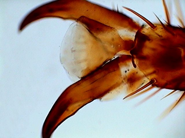
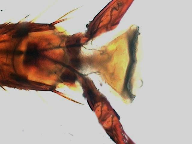
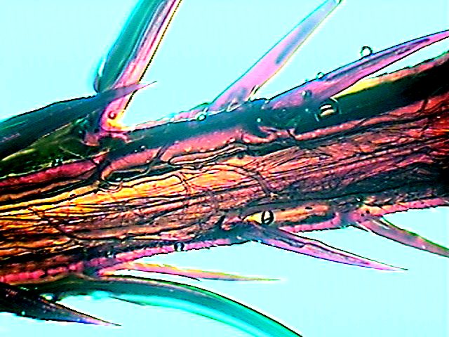
From upper left going clockwise: claw
of the pretarsus in lateral view; pretarsus in dorsal
view; a ventral view of the pretarsus. The three pictures show clearly
the arolium,
an adhesive cup that allows cockroaches to climb vertical walls, even
polished ones. The fourth picture shows through the translucent
chitinous
skeleton the dense net of tracheae that oxygenates the
powerful leg muscles.
|
If
it is
desired to learn the principles of parasitological investigation,
an important
branch of general biology, agriculture, veterinary medicine and
human medicine, dissection is an essential practice. But you must
master the
correct
techniques.
In schools the use of real dissections (the specimens usually adopted are frogs,
certain large
locusts
and cockroaches) can perhaps be
replaced with great
disadvantage of course by the use of virtual reality software or more
modestly with
three dimensional images of some good dissections, or
with drawings, based on the same source. This could probably save
thousands of specimens used annually in schools.
But
it
cannot be forgotten that the anatomy of the animals thus represented,
which
must necessarily be taught to the students, was discovered by
scientists who
made the necessary dissections and published the corresponding
documentation.
If
Leonardo
da Vinci and Vesalio had not dissected human beings, we would
perhaps still believe that the veins and arteries (like the tracheas of
the
insects), were
empty tubes that transported air from the lungs to body tissues. And
without having
learnt to dissect, no surgeon could save lives via a surgical
operation.
In
fact we
hope that some of the images presented here can be used with that aim.
Aside from
the images of the University of Alberta's website, the only dissection
of a
cockroach (in
English) extensively mentioned on the internet is a promotion of a
video tape, and is illustrated online only with
small
images, several of which are semi-concealed by a band of lines.
|
|
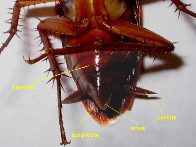
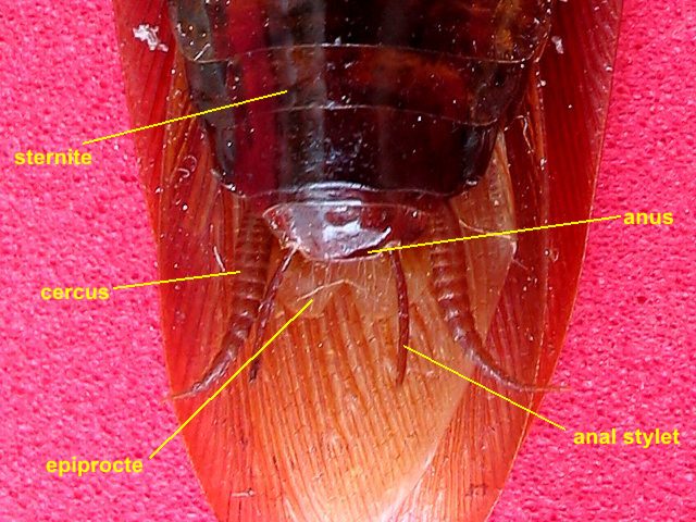
Female
abdomen: note the acute end, the two sensitive cerci, and the short
wings.
|
Abdomen,
male: note the rounded end, the two anal stylets and the long wings.
|
The
dissection
Now we have the information
we
need to proceed with our cockroach dissection.
For
one
complete, detailed and magnificently informed dissection, see the
Canadian website
referred to previously.
Of
course microscopists
will not lose the opportunity to study the pretarsus of the legs that
contains
the secret of why cockroaches climb vertical walls, the structure of
the
trachea with its characteristic chitinous spiral support, the abundant
fatty tissue
in the abdomen, the structure of the compound eye of an insect with top
lighting, the wings and antennae, etc.
And,
if a microscopist
has hypochlorite, or even better sodium or potassium hydroxide, they
may want to
prepare
the mouthparts of the insect.
But
here we
will only give the directions to separate the alimentary canal, from
which we
will extract the parasites.
According
to the interests of the
investigator the
dissection can be made dorsally or ventrally. The ventral way is the
easiest if
you want to study the intestinal fauna, so it is the one to be used
here.
Of
course
if a representative research is to be conducted, enough male and
female
cockroaches, as well as nymphs, must be dissected to give a
statistically
significant sample.
But
for
this article only a few adults of both sexes are used.
Some
authors assign to the adults of P. americana a length of up to 44 mm. In Cancún and Durango I have only been able to measure
(on some tens of individuals collected) a maximum body length of 35 mm. Ten
millimetres are
significant when making such small insect dissections.
A list of
the parasites I have found is:
COMMENSALS
|
|
amoebae
|
Endamoeba blattae
|
ciliates
|
Nyctotherus ovalis
|
PROBABLY
PARASITES
|
nematodes
|
Hammersmidtiella diesingi
|
|
Thelastoma bulhoesi
|
These species will be described in the
following parts of this series. I heartly acknowledge the help of
Professor Nora
B. Camino, helminthologist at the University of La Plata, La Plata
city,
Bs.As., República Argentina, for the confirmation of the
nematode genera to be described here.
APPENDIX
Cockroach control
If they are not a real nuisance, and if their
activities are limited to the garden or the wild, leave them alone. In
nature they
have an important role in the disposal of ecosystem detritus. Use all
your best
efforts not to offer them a way into your house. A clean house,
without many crevices, and without poorly stored food, rarely
suffers from large cockroach invasions. Attempt to control them only if
their populations increase beyond that of some occasional visible
visits.
The oldest and most efficient method is
the use of boric acid crystal powder at sites near the
places were
cockroach retire and procreate. They eat the sharp crystals which
lacerate and
cause bleeding of the gut.
Boric
acid bait
Note:
do not
breathe the powder, use rubber gloves to prepare the bait.
- One half cup
(350 grams) boric acid - looks like white powder and is
available in pharmacies
- one cup flour
- two heaped
spoonfuls of sugar
- water
Mix
boric acid, flour and sugar.
Moisten with just enough water to make a paste that can be formed into
small cylinders or balls.
Make as many of them as you can. Distribute them in critical concealed
places.
Nowadays, with exactly the same rationale is the use of
ground diatomaceous earth; not the powder for domestic
pool filters, but
the type sold by many garden centres to control insect pests.
Broken
diatoms
have very sharp silica fragments.
If this seems cruel, think of the
insects (cockroaches and who knows what else) inmobilized by neurotoxic
chemicals, condemned to die of
dehydration
and starvation. And of the collateral effects of this type of
insecticide on
the whole ecosystem. Such methods kill not only cockroaches, but
any
insect within its reach, and has accumulative and deleterious effects
on
birds and rodents that eat the carcasses.
|
