Micscape: Article of the month (Nov 95)
Observations on Two Rare Desmids From N.W.
Scotland, British Isles.
by
William Ells
Coniferae, Walnut Tree Lane, Loose, Maidstone,
Kent.ME15 9RG. England.
15 September 1995
Desmids are an attractive unicellular microscopic form of
green algae (Chlorophyta) found only in freshwater. They belong
to the class Zygnemaphyceae which is unique among the freshwater
algae in that each plant can join with another of the same
species in a process known as conjugation to form zygospores.
Although zygospores have not been found for all species.
Method:
Samples are collected from boggy areas or the margins of Lochs in
North West Scotland. Part of the sample is preserved in about 5%
formalin the remainder is examined in the living state over a
period of several days. I prefer to examine live, not only is the
bright often vibrant green attractive, it makes them easier to
pick out, particularly the smaller species, from amongst any
debris. Also they can often be observed multiplying asexually by
division. and more rarely conjugating to form zygospores, they
normally do this in adverse conditions usually when the habitat
is drying out, forming new plants when climatic conditions are
right. Attack by fungi or fauna can also be observed, Brook &
Ells (1987). The drawback of examining live material is that
drawing or photography is much more difficult owing to movement
in the water caused by the fauna or the temporary mount drying
out.
All observations were made with a Nikon Skt. trinocular
microscope using 20:1 plan achromatic and 40:1 plan fluorite
objectives, eyepiece HKW 10:1. Photographs were taken with A
Praktica SLR. using a Zeiss projection eyepiece 4:1. Drawings
were made using a Biolam camera lucida.
Observations:
A sample was received from Loch Chealamy, Strathnaver, Sutherland
on 28.9.94 and examined the same day, this was found to contain
some 50 species of desmid including Closterium nematodes
Josh. var. proboscideum W.B.Turner 1892. This is the first
time the species had been found in a living sample from the
British Isles, the type C.nematodes var. nematodes
has not been seen. Brook (personal communication) also examined a
sample and confirmed the species and variety.
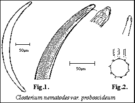
 The cells (Fig.1.) are 8-12
times longer than Broad, moderately curved. The walls have a
thickened ring just behind the apices which make this species
distinctive and gives rise to the name nematodes, 5-9
broad ridges known as costae can be seen across the breadth of
the cell these run from apice to apice, when the two edges of the
costae are brought into focus where they meet the cell wall they
are seen as pairs of lines, the cells are circular in the centre
with the coastae spaced evenly around the cell wall. There are
5-7 pyrenoids in each semi-cell.
Tell & Couté 1993, showed the shape of the costae of Closterium
costatum Corda ex Ralfs 1848. in Scanning Electron
photomicrographs. Fig.2. showing how the centre of a cell
in cross section should look is based on these micrographs.
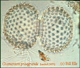
Cosmarium praegrande Lundell (1871). Several specimens
were found in a sample from Rhiconich, Portle Vorchy, Sutherland
in October 1993, This large species of Cosmarium is
exceedingly rare in Britain. The dimensions of the Rhiconich
specimens; Length 120 microns. Breadth 71 microns isthmus 26.5
microns; are larger than those described by W.&.G.S.West
(1908). Also larger than those described and figured by Prescott
et.al.(1981).
 The semi-cells are spheroid, end view
circular, the cell walls are densely covered with prominent
conical granules (or warts) except for a small area at the apex
and finely punctate between the granules. The size of the
granules are as those figured by W.&.G.S.West. (after
Lundell) those figured by Prescott et al. are smaller.
The specimen in the photomicrographs has had a neat hole cut
in the wall of one semi-cell, the piece removed can be seen
nearby, the hyphae of a microscopic fungi can also be seen, the
cell is devoid of any vegetable matter.
Acknowledgements: Thanks to Mr.Alan Joyce of Sutherland
for the samples from Chealamy & Rhiconich, to Dr.J.W.G.Lund
CBE,DSc,CBiol,FRS. Freshwater Biological Association, England,
for figures from the Fritsch Collection of Algal Illustrations.
and to Prof.A.J.Brook PhD,DSc,FRSE. of The University of
Buckingham for his comments on the sample from Loch Chealamy.
Comments to Bill
Ells welcomed.
References:
Brook A.J. & Ells W. (1987) The feeding of Amoeba on
Desmids. The Quekett Journal of Microscopy Vol.35. Part 7.
Prescott G.W., Croasdale H.T., Vinyard W.C., &. Bicudo De
M. C.E. (1981) A Synopsis of North American Desmids. Part
2. section 3. University of Nebraska Press.
West W.&.G.S. (1908) A Monograph of the British
Desmidiaceae. Vol. 3. The Ray Society.
Tell G. & Couté A. Cryptogamie Algol 1983. 14(1)
43-63.
© Microscopy UK or their
contributors.
Please report any Web problems
or offer general comments to the Micscape Editor,
via the contact on current Micscape Index.
Micscape is the on-line monthly
magazine of the Microscopy UK web
site at Microscopy-UK
WIDTH=1
© Onview.net Ltd, Microscopy-UK, and all contributors 1995 onwards. All rights
reserved. Main site is at www.microscopy-uk.org.uk with full mirror at www.microscopy-uk.net.
 The cells (Fig.1.) are 8-12
times longer than Broad, moderately curved. The walls have a
thickened ring just behind the apices which make this species
distinctive and gives rise to the name nematodes, 5-9
broad ridges known as costae can be seen across the breadth of
the cell these run from apice to apice, when the two edges of the
costae are brought into focus where they meet the cell wall they
are seen as pairs of lines, the cells are circular in the centre
with the coastae spaced evenly around the cell wall. There are
5-7 pyrenoids in each semi-cell.
  The semi-cells are spheroid, end view
circular, the cell walls are densely covered with prominent
conical granules (or warts) except for a small area at the apex
and finely punctate between the granules. The size of the
granules are as those figured by W.&.G.S.West. (after
Lundell) those figured by Prescott et al. are smaller.