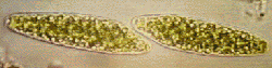
 Fig 1. Shows, in
transmitted light without stain, recently divided cells of the
rare saccoderm desmid Spirotaenia alpina Schmidle (1895).
New to the British Isles, Brook 1992.
Fig 1. Shows, in
transmitted light without stain, recently divided cells of the
rare saccoderm desmid Spirotaenia alpina Schmidle (1895).
New to the British Isles, Brook 1992.
Desmids a microscopic form of freshwater algae, exude mucilage, a
transparent or almost transparent gelatinous substance, sometimes
in copious quantities that can be easily seen under a microscope
using normal transmitted light, for example in the desmid genus Hyalotheca
which forms long filaments. In other genera it may be only
faintly seen, if at all, and phase contrast or some method of
staining will have to be used to show the extent of the mucilage.
In my article (Micscape Dec.95) Some Observations on Freshwater Algae I referred to using Alcian blue in my experiments on Astrococcus the mucilage envelope of which did not not absorb the stain, although desmid mucilage always does. I also mentioned the use of diluted Indian ink the method shown here. A little diluted ink is placed on the slide next to the cover glass where it may spread under by capillary attraction or can be drawn under by removing water from the other side of the cover with blotting paper.
Fig 1. Shows, in
transmitted light without stain, recently divided cells of the
rare saccoderm desmid Spirotaenia alpina Schmidle (1895).
New to the British Isles, Brook 1992.
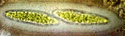 Fig 2. With Indian ink
added shows the mucilage envelope surrounding the cells. Size of
cells 71.5 um long; 18.5 um. broad. In fig.1. the distinctive red
ends to the chloroplast, can just be seen in the older apices,
those furthest apart. These appear in both apices of fully mature
cells.
Fig 2. With Indian ink
added shows the mucilage envelope surrounding the cells. Size of
cells 71.5 um long; 18.5 um. broad. In fig.1. the distinctive red
ends to the chloroplast, can just be seen in the older apices,
those furthest apart. These appear in both apices of fully mature
cells.
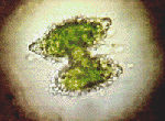 Fig 3. Shows the much
more dense mucilage surrounding a species of the placoderm desmid
Staurastrum, mucilage can actually be seen being expressed
from pores in the cell wall.
Fig 3. Shows the much
more dense mucilage surrounding a species of the placoderm desmid
Staurastrum, mucilage can actually be seen being expressed
from pores in the cell wall.
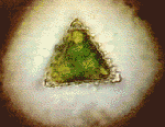 Fig 4. shows the end
view of the same desmid, size 47um. from base to apex.
Fig 4. shows the end
view of the same desmid, size 47um. from base to apex.
My next article will show desmids viewed by polarised light.
Reference:-
Brook A.J. (1992) Spirotaenia alpina (Zygnemaphyceae: Mesotaeniaceae), a Saccoderm Desmid New to the British Isles: Revised Description and Observations on Cell Division. British Phycological Journal. 27. 1.March 1992.
Comments to Bill Ells welcomed.
Acknowledgement
The Micscape Editors thank William Ells for contributing this article.
Please report any Web problems
or offer general comments to the Micscape Editor,
via the contact on current Micscape Index.
Micscape is the on-line monthly
magazine of the Microscopy UK web
site at Microscopy-UK