MICSCAPE ARTICLE
Some Observations on Freshwater Algae
by
William Ells
Coniferae, Walnut Tree Lane, Loose, Maidstone, Kent.
ME15 9RG. England.
13th December 1995
What is loosely known as pond life, (freshwater life is a
better term), especially the microscopic flora & fauna, can
provide enough interest to last a lifetime. My main interest is
in the algae, particularly the desmids. The best way to study
algae, with the exception of the diatoms, is to examine living
material. Algae fixed and made into fluid mounts can be useful
for reference purposes, but unlike the diatoms which can be
cleaned and mounted in a resin mountant, fluid mounts are rarely
100% permanent.
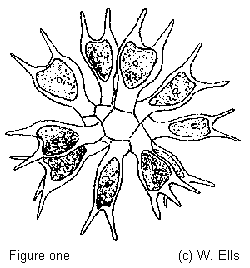 Although I study desmids, now and again
another alga of special interest is found. In 1980 one of my
samples had a coenobium (a colony of unicellular organisms) of Sorastrum
americanum [Bohlin] Smidle, a member of the Chlorophyta
(green algae) which I identified from Prescott (1954). My Fig.1.
is re-drawn from his figure. I thought little of this until
another coenobium turned up in a sample from Sutherland,
N.W.Scotland in 1992. Realising this was only the second colony I
had seen in twelve years, I looked it up in West & Fritsch
(1904-1927) not available to me in 1980 when the first specimen
was seen. They state 'it occurs in the Outer Hebrides' no figure
of this species is shown and no mention of it being found in
mainland Britain. Lund (personal communication) informs me he has
no records of S.americanum for England or Wales since West
& West (1905), although it is known from Africa, America
& Europe. It is more common in the tropics than in the
temperate zone where it is very rare. This is where an amateur
like myself can make a useful contribution to the knowledge of
algae by recording the occurrence and distribution of rare
species at The Freshwater Biological Association, Windermere,
England.
Although I study desmids, now and again
another alga of special interest is found. In 1980 one of my
samples had a coenobium (a colony of unicellular organisms) of Sorastrum
americanum [Bohlin] Smidle, a member of the Chlorophyta
(green algae) which I identified from Prescott (1954). My Fig.1.
is re-drawn from his figure. I thought little of this until
another coenobium turned up in a sample from Sutherland,
N.W.Scotland in 1992. Realising this was only the second colony I
had seen in twelve years, I looked it up in West & Fritsch
(1904-1927) not available to me in 1980 when the first specimen
was seen. They state 'it occurs in the Outer Hebrides' no figure
of this species is shown and no mention of it being found in
mainland Britain. Lund (personal communication) informs me he has
no records of S.americanum for England or Wales since West
& West (1905), although it is known from Africa, America
& Europe. It is more common in the tropics than in the
temperate zone where it is very rare. This is where an amateur
like myself can make a useful contribution to the knowledge of
algae by recording the occurrence and distribution of rare
species at The Freshwater Biological Association, Windermere,
England.
Sorastrum americanum is quite distinctive although it
bears a superficial resemblance to some Pediastrum species
i.e. the colony is only loosely attached, not compact, and it is
globose not flat like Pediastrum. There are commonly 16-64
heart shaped cells; there are out of focus cells coming forward
and away from the focal plane in the figure (Fig.1.). The only
other British species S.spinulosum Naeg. has bean shaped
cells with small processes, described by West & West as a
rare species found in boggy pools etc. Lund however states 'S.spinulosum
is cosmopolitan, somewhat common here since it is recorded from
several places in England, Ireland & Scotland though not,
apparently, recently.'
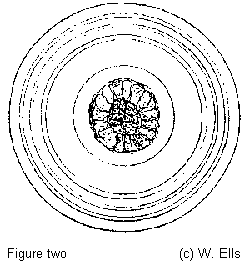 Another alga I found interesting, very
common in Britain frequently found in boggy pools where desmids
are abundant, is Asterococcus superbus [Cienk] Scherffel
(1908) =(Pleurococcus superbus Cienk; Glococytis
infusionum W.& G.S.West) The only British species, it has
large cells, single or in groups up to 8, 25-30 microns in
diameter, and a striking lamellated mucilage envelope often
reaching 180 microns in diameter the concentric rings of which
first attracted me to the species. (It has a nice species name I
can actually pronounce, I am still looking for another, the
desmid Cosmarium superbum so far only recorded from a few
sites in North America, Prescott et.al.[1981]). The rings can
often be seen under the microscope in normal transmitted light,
but not as clearly defined as shown in Fig.2. drawn from life the
rings have been over emphasised for reproduction in this article.
With some specimens you may find the only way the rings can be
seen is by staining or using phase contrast. With my phase 'DM'
type the rings appear white on a grey background.
Another alga I found interesting, very
common in Britain frequently found in boggy pools where desmids
are abundant, is Asterococcus superbus [Cienk] Scherffel
(1908) =(Pleurococcus superbus Cienk; Glococytis
infusionum W.& G.S.West) The only British species, it has
large cells, single or in groups up to 8, 25-30 microns in
diameter, and a striking lamellated mucilage envelope often
reaching 180 microns in diameter the concentric rings of which
first attracted me to the species. (It has a nice species name I
can actually pronounce, I am still looking for another, the
desmid Cosmarium superbum so far only recorded from a few
sites in North America, Prescott et.al.[1981]). The rings can
often be seen under the microscope in normal transmitted light,
but not as clearly defined as shown in Fig.2. drawn from life the
rings have been over emphasised for reproduction in this article.
With some specimens you may find the only way the rings can be
seen is by staining or using phase contrast. With my phase 'DM'
type the rings appear white on a grey background.
I experimented by adding diluted black Indian ink to a sample
on a slide, the mucilage does not take up the ink, thus showing
up white on a dark grey background. To a specimen on another
slide I added Alcian blue. The mucilage, invisible or almost so,
that surrounds or is extruded by most desmids will absorb this
stain and be revealed. The Astrococcus mucilage must be
different or it has a skin or pellicle preventing the stain being
absorbed, only the outer ring stained and became easier to see.
To yet another slide I added Lugols iodine diluted to the
colour of pale sherry, (sometimes used as an algae preservative).
This must have penetrated the mucilage envelope, because the
chloroplast of the cell turned almost black, deep purple usually
indicates the presence of starch, the mucilage was not coloured
by the iodine. NOTE the iodine, stains etc. were added by placing
a few drops around the cover glass, these spread through the
sample by capillary attraction.
Finally I tried to see the starch grains using polarised light
and a 100:1 oil immersion objective. This failed, probably
because the lamp a 6 volt 15 watt; ordinary tungsten not halogen
on my microscope, a Nikon Skt. is not powerful enough to cope
with crossed polars at this high magnification.
West & Fritsch state this organism has an eye spot and a
motile phase although neither was seen during my observation.
This article first appeared in Balsam Post The Newsletter of
the Postal Microscopical Society (British) 1992. It has been
revised for the W.W.W.
Acknowledgements
Thanks are due to Mr Alan Joyce for the samples from Scotland,
and to Dr J.W.Lund CBE.DSc.CBiol.FRS. of The Freshwater
Biological Association, Windermere, England, for his information
on Algae records and figures from the Fritsch Collection of Algal
Illustrations.
References
Prescott G.W. (1954) How to Know the Freshwater Algae.
Wm.C.Brown Co.Dubuque, Iowa.
Prescott G.W. Croasdale H.T. Vinyard W.C. & Bicudo C.De M.
(1981) A Synopsis of North American Desmids Pt. 2. Section
3. University of Nebraska Press.
West G.S. (1904) revised Fritsch F.E. (1927) A Treatise on
the British Freshwater Algae. Cambridge University Press.
London.
Comments to Bill
Ells welcomed.
© Microscopy UK or their
contributors.
Please report any Web problems
or offer general comments to the Micscape Editor,
via the contact on current Micscape Index.
Micscape is the on-line monthly
magazine of the Microscopy UK web
site at Microscopy-UK
WIDTH=1
© Onview.net Ltd, Microscopy-UK, and all contributors 1995 onwards. All rights
reserved. Main site is at www.microscopy-uk.org.uk with full mirror at www.microscopy-uk.net.