My brother Ian and I recently treated ourselves to a boxed mixed selection of 19thC papered slides. Some slides were of quite thick parts of insects so offered an opportunity to explore the use of near infrared which has a higher transmittance through the exoskeleton than visible. I also wished to compare the NIR results with just using deep red filters. To date for NIR I have used either a home modified Sony S75 3 Mpixel consumer digicam with fixed 3X optical zoom lens or a now aging 1 Mpixel monochrome camera that is used for 'diatom dotting' type studies. I had recently upgraded the latter to a 6 Mpixel ZWO ASI178MM model but had not yet tried for NIR.
A selection of slides are below comparing brightfield with deep red and NIR. For brightfield a NIR blocking filter such as the typical CM500S needs to be used so that only capturing a visible image. Consumer digicams always have this sky blue filter over the sensor. For a deep red at 685 nm I use an unmounted sheet from Knight Optical 685FAP5050. Near IR used three mounted 52 mm camera lens filters. The Hoya R72 typically used for landscape NIR studies and a 760 nm and 850 nm. The latter two were the 'dHD' Chinese made brands and quite good value on eBay.
The ZWO camera has a 1/1.8" sensor with 14 bit ADC. A small sensor is useful on my Nikon E800 and Zeiss Photomicroscope III stands for its main role of 'diatom dotting' as the cropped visible field is useful for frustule detail to fill the sensor frame. However, for low mag studies of large subjects, in this case using a Nikon 2X / 0.10 planapo, the sensor only captures a small part of the field. A 0.5X relay lens was to hand but required the camera to be used in an eyepiece rather than the vertical photo port as shown below.
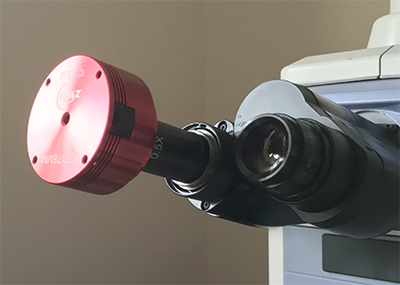
The 0.5X relay lens has an eyepiece fitting so unsuited for the photo-port with C-mount adaptor.
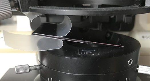
To protect the plastic 685 nm deep red filter the protective film is just peeled back when using on the microscope (sitting on the Nikon Eclipse de Senarmont DIC module).
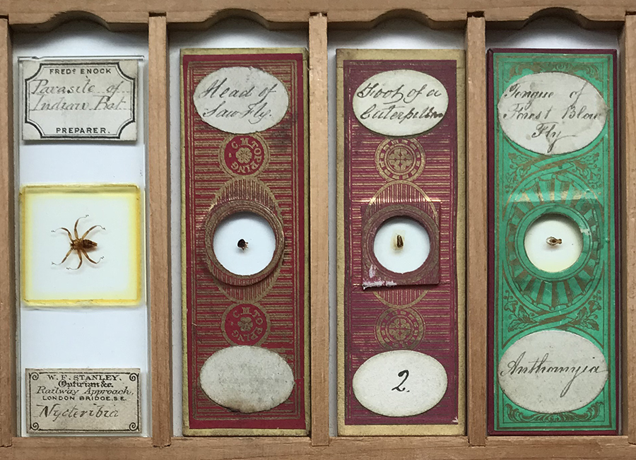
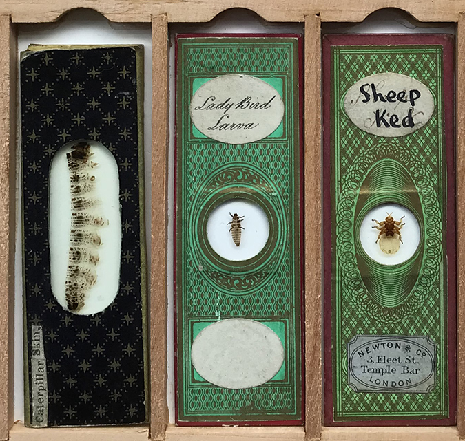
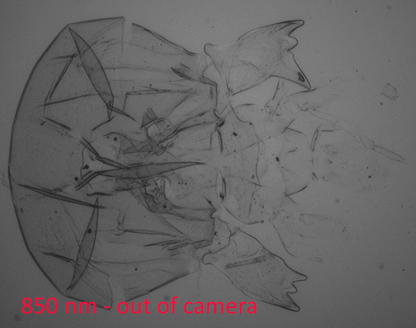
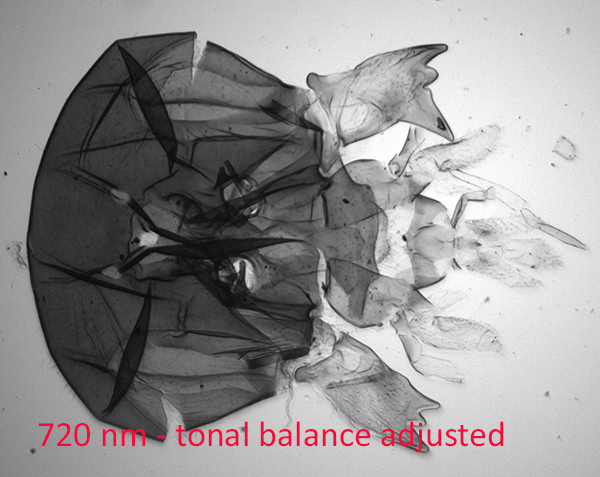
Lefthand images below are in brightfield, others as labelled. Deep red and NIR images out of camera are tonally flat and become increasingly so at longer wavelengths. The tonal balances were adjusted to create a better contrast B&W image.
Using the 685 nm deep red filter
I was surprised how effective just using a deep red filter can be rather than using near IR below 700 nm for many slides where the exoskeleton was not too thick.
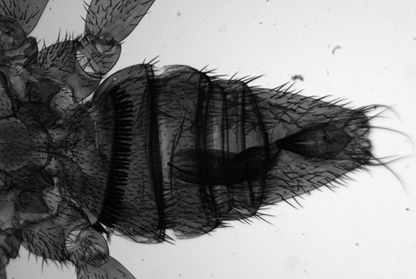
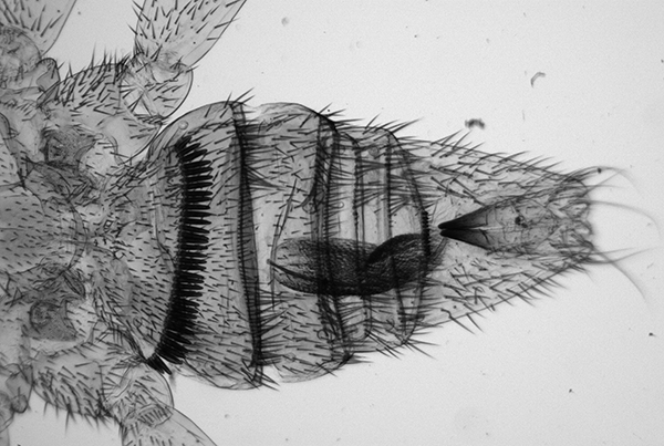
Abdomen of the parasite of an Indian bat. Righthand image 685 nm.
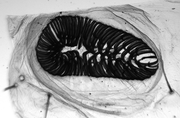
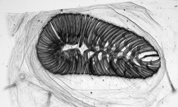
Foot of a caterpillar. Righthand image 685 nm.
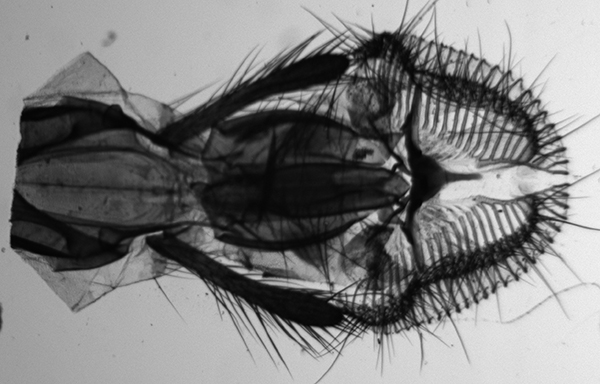
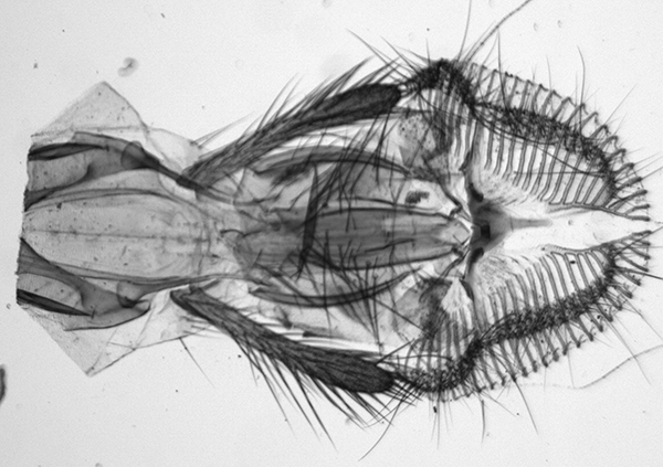
Tongue of forest blow fly Anthomyia. Righthand image 685 nm.
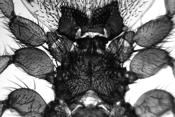
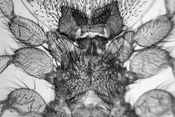
Sheep ked abdomen. Righthand image 685 nm.
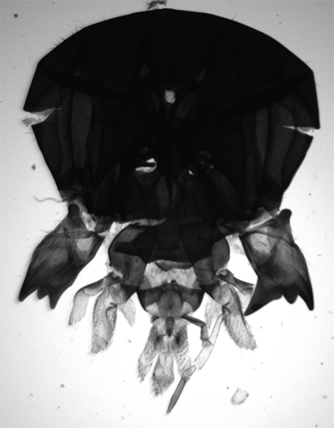
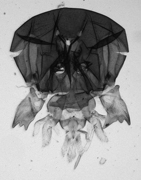
Head of sawfly. Righthand image 685 nm. When the exoskeleton becomes thicker, a deep red is less effective but still offers some increase in transmittance.
The 685 nm image shows the increased depth of field.
Using near IR filters
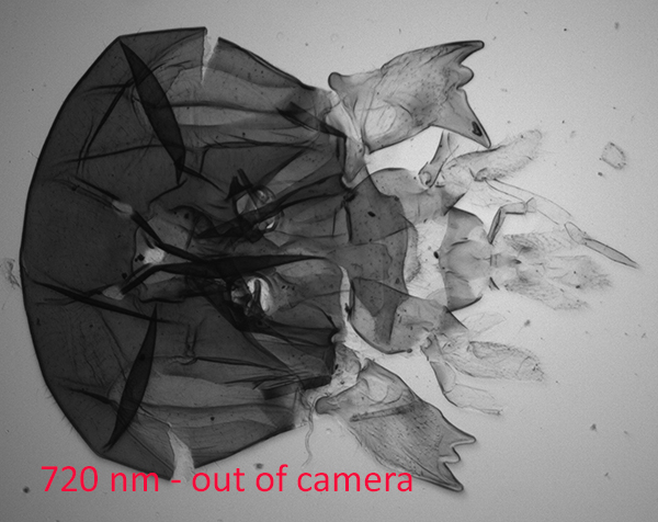
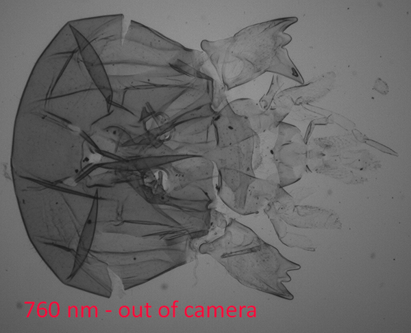
The sequence below shows the sawfly imaged with increasing wavelength, i.e. moving from deep red into true near IR greater than 700 nm. They can be compared with the brightfield and deep red 685 nm images above. For each subject the optimal wavelength required can vary. The denser parts reveal more detail but thinner more transparent areas seen in brightfield become washed out lowering the overall tonal range which is hard to recover with post capture editing. In this case 720 nm near optimal for good contrast after tonal balance correction.
Very even illumination at mags as low as 2X can be tricky even with a modern stand like the Nikon Eclipse E800. Near IR seems to reveal any unevenness more than brightfield visually. A tungsten filament of course has a large near IR component in its black body spectrum.
Comments to the author David Walker are welcomed.