This article is about three of my favorite protozoans. I will be presenting you with some new images of each which I just took in the past few weeks. I will provide a brief commentary (well, brief, for me). These images were taken of live organisms and I used a Nikon SMZ-1000 Stereo Microscope with a Jenoptik Pro Camera which is tethered to an ASUS laptop computer.
Why a stereo microscope and not a compound? Well, if you’ve ever tried to photograph and of these organisms live, you will soon discover that they are generally very active and overall do not respond well to anesthetic procedures and even attempts to slow them down by using such solutions as Methocel can produce distortion and the additional viscosity can create problems with resolution. Even using a stereo system, considerable patience is required.
What are the three organisms? Well, let’s begin with the micro-whales as I like to call Spirostomum. In relation to, Paramecium, for example, which averages about 250 microns, this is indeed a “whale”. The images of this one are the least satisfactory, in large part, due to the extraordinary contractility of this large protozoan. Spirostomum ambiguum can be over 5,000 microns in length and has the fastest contractility rate of virtually any organism known. When disturbed or threatened it suddenly reduces itself to about 1/3 its full length. This species has a moniloform or “beaded” macronucleus and a very large posterior contractile vacuole. Unfortunately, the batch which I was dealing with was singularly uncooperative and I can show you a partially extended specimen and a couple of closeups. However, from a previous batch a few years ago using a different microscope, I do have a couple of images showing the remarkable length and the large size of the contractile vacuole.


In the more recent images, the first one shows some of the fibrils that run through the organism and are part of the mechanism involved in the contractility.

The next image is of a contracted form and shows the entire surface covered with cilia. We also get hints of facets of the “beaded” macronucleus.

In this closeup of a portion of the organism, you can see more clearly a part of the macronucleus.

Next, let’s take a look at Stentor coeruleus, distinctive because of its blue-green pigment. I’ll never forget my first encounter with the marvelous trumpet-shaped Stentor coeruleus. I was in my early teens and had been collecting some water samples from Antelope Creek which was in a nearby park. The name comes from the Achaean warrior who was in the battle at Troy and was reputed to have an incredibly loud and forceful voice, apparently reminding some of ancients of the sound of a slapinx or trumpet. Such an instrument had no valves and was quite long and yet relatively light. The two examples below are from Greek pottery.


The figures depicted look quite young. About half of those born in ancient Greece died before reaching adolescence and the average life expectancy was about 35. However, there is not a great deal of agreement and the estimates are derived from ancient sources which are not reliable by modern standards. Other estimates put the average life span during the Roman Empire at a mere 25 years and 33 during the Middle Ages. In ancient Egypt the estimates are even shorter with males living only about 23 years and females 33 years. Interestingly, almost all statistics, including modern ones, show women as living several years longer than males. In 2021, the estimate for males worldwide is slight over 73 and for females, nearly 80. So much for the myth of the weaker sex.
A depiction of a Roman warrior with a trumpet suggests someone older than one would expect from the numbers.

In addition, we have the Medieval or Heraldic trumpet which typically had a banner attached to it. Here are two examples, the second with an escutcheon showing a lion rampant.


Finally, there is the Renaissance trumpet. This particular example is a rather plain, but nicely crafted, copper one. I suspect, however, that these often were fitted with hooks to hold banners, since it’s hard to imagine that the Borgias and Medicis wouldn’t take every opportunity to advertise themselves.

Now, in exasperation, you are probably yelling, asking what all of this has to do with Stentor. I could go on about Flügelhorns, French horns, trombones, English horns, etc., but I’ll restrain myself. Well, I warned you that I love tangents, (and my wife loves tan gents), but your patience will be rewarded, for remarkably, Stentor does have a significant resemblance to some of these types of trumpets and you will see. First up is a view of two Stentor nicely extended and the color suggests a fine copper patina on the trumpet. Interestingly, this pigment is exclusive to Stentor and has been given the name of stentorin. I guess these clever protists got together and patented it.

In the next view, you see three specimens, two of which are curving, showing how flexible they can be. The organisms are attached to bits of debris, which is likely composed of huge numbers of bacteria. The Paramecia appear to be feeding on that material. Stentor have “holdfasts” which allow them to attach and then detach when disturbed or when food is exhausted in their particular area. It is not usual to see Stentor swimming freely in a somewhat contracted state.

Sometimes, they even attach to each other.

Now, let’s take a closer look at a single individual and see what we can discover. First, a general view which shows us an extended organism with its posterior anchored in what is likely bacterial detritus and then displaying it’s trumpet-like form as it extends up to a large opening which is the cytostome. As we shall see this is a rather complex structure. Notice also that the stentorin pigment is readily discernible.

If we invert the image, we get a slightly better sense of the cystostome.

Now, if we increase the magnification of the regular image just slightly, we can see, if we look carefully, that the cystostome is ringed by cilia.

Upon converting the image to monochrome, we can see a swirl in the form of the cystostome. This is of special interest, because when we observe an active feeding specimen, we can see that the cilia produce a motion that brings particles down into the swirling structure which then carries them further down into a vacuole which when full, separates and moves down into the body in order to digest the content. However, some particles are immediately rejected as unsuitable as food and are swirled out and not moved into the vacuole.

A closeup of the area of the cystostome reveals that this gives a further indication of the complexity of this structure.

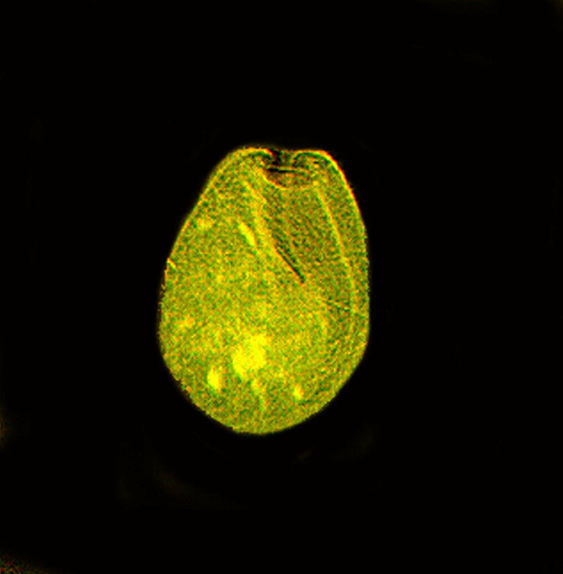
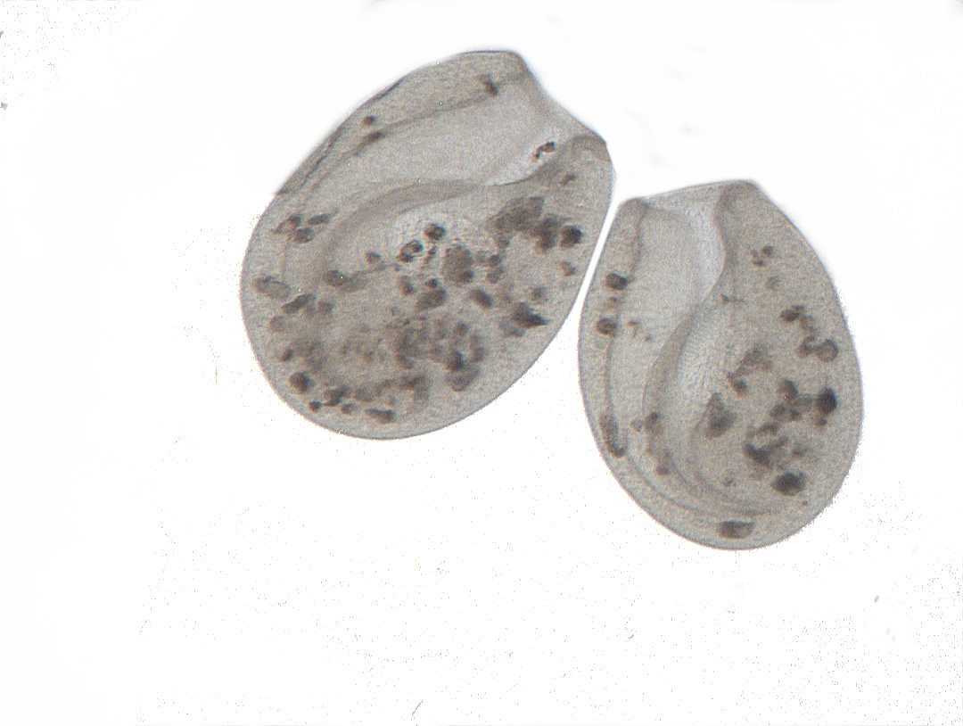
O.K., now let’s move on to what may well be my favorite of these three protists, namely, Bursaria truncatella. The way it glides around on the slide is nothing less that elegant; also frustrating when one is trying to photograph it. I have described it as a vase and I feel that even more vividly watching it in motion. Some may well feel that this description is a bit of a stretch, but I’ll stick with it. First I’ll show you an image of an art glass vase and then a Bursaria and you can make your own comparisons. If you come up with a description you like better than “vase” and would like to share it, that would be great.


B. truncatella can be slightly over 1000 microns and it has an extraordinarily large cytostome or mouth which curves down from the anterior end to the very tip of the posterior end. The anterior end is also flexible and so it can engulf rather large prey whole. They feed ravenously on Paramecia which they ingest entire. The macronucleus is a long, thin, band-shaped affair which curls around the cell in an unpredictable manner. There are numerous micro-nuclei.
I am going to show you a few images using several different graphics techniques to reveal, I hope, some detail that might otherwise not be noticed.
This first image is brightfield where I have created a white background to increase the contrast.

I then took this image and used the invert function on it. I think you will agree that the long curved cytostome is more distinctive in this image.

Perhaps equally effective is the application of the enhanced lighting function to the inverted images whereby the color is slightly altered.

Finally, another brightfield image followed by the application of the invert function to it. The brightfield image is not as sharp as I would like and this is a very challenging organism to photograph.

However, by using the invert function, the image is a bit sharper in that it reveals somewhat more detail.

Also, with a bit of patience and perseverance, one can sometimes luck out and get some sharper images. I was fortunate enough to have that happen and will show you three more images each having two specimens.

This next one shows them near a mass of bacterial detritus.

The final image is where the two and in very close proximity and it’s not possible to tell if the they are canoodling, attacking, or just accidentally bumping into each other.

I hope you have enjoyed this brief look at these three intriguing protists and that you will have good fortune in finding them and studying them for yourselves.