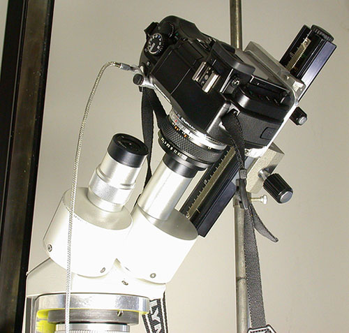
Figure 1 (Click image to view master)
|
Using an Olympus E-330 DSLR with a Binocular Stereomicroscope by Ted
Clarke, USA |
My earlier
Micscape article, “A Gun-Mount for Nature Photomacrography”,
gave an introduction to my use of this very innovative camera. A
subsequent article in Microscopy
Today, “Olympus E330 DSLR for
Photomicrography with Older Design Microscopes”, demonstrates
how this camera can be used with an Olympus 28 mm f/2.8 camera lens
held over a 10X eyepiece to record a full 18 mm FN without vignetting
even though the eyepiece is not high eyepoint. The camera was mounted
in my rigid macro stand to avoid blurring from shutter vibration,
which has been my practice when using 35 mm SLR camera backs for
photomicrography and photomacrography. I had planned to follow this
with evaluation of the E-330 for scanning light photomacrography. My
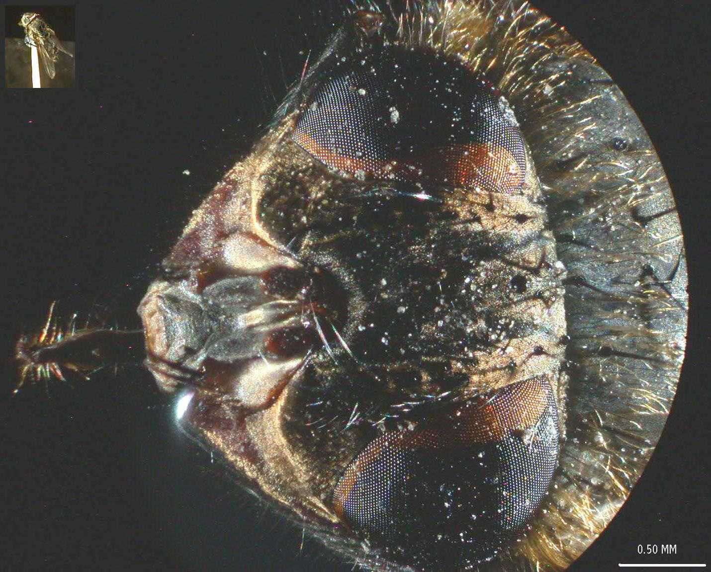
Modern Microscopy article on scanning light contains Photo 1 showing
the head of a housefly recorded with my Nikon Coolpix 995 mounted to
a 10X high eyepoint eyepiece in the bellows system. The E-330 will
allow direct projection of the macro lens image onto the camera
sensor without the need for an eyepiece and with twice the pixel
count for a larger field size at the same spatial resolution. I have
also been working with a home-made transmitted light illuminator for
use with my stereomicroscope so I can look at lake water organisms
with the grandkids. I wondered whether there was a way for me to
use my E-330 for digital images from the binocular Meiji
stereomicroscope used with the new illuminator.
Figure 1 (Click image to view master)
I
suspected that it might be possible to mount the E-330 on the bellows
rail of one of my pair of home-made fiber-optic double condenser
illuminators described in my Microscopy Today article “Brightfield
Illumination of Large Field Sizes”. These illuminators use an
Olympus Auto Bellows as a focusing rail for their components. My
previous experience with the crack-prone plastic inserts used in the
Olympus Auto Bellows is described in my Microscopy Today article (p.1,
p.2)
“Heavy Duty Camera Bellows for Digital Imaging”. The
Kodak MegaPlus 1.6i digital camera caused cracks to form in the
female dovetail inserts. These were noticed before total failure
occurred allowing the expensive camera to fall. Even though the
double condenser application for the Auto Bellows does not impose the
heavy loading of the digital camera, cracks were found in dovetail
mounts after about 15 years of use. I replace all of the plastic
inserts with precision machined zinc alloy ZA-12 inserts made in my
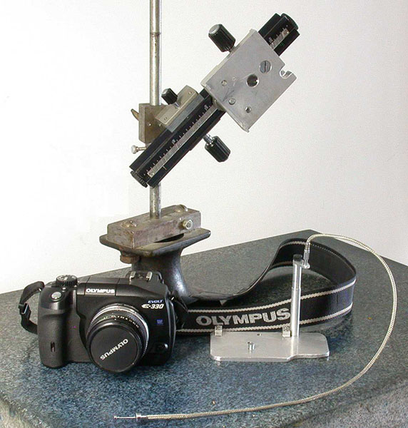
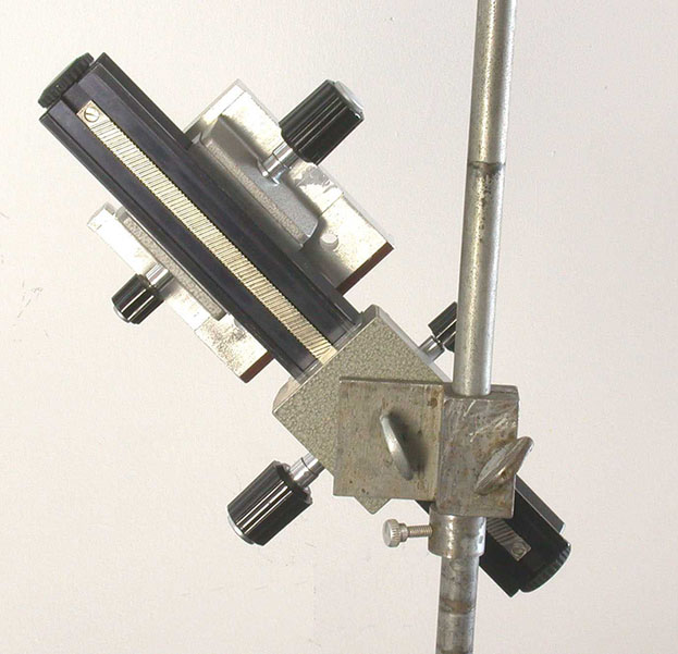
home shop. Photos 2 & 3 show the front and back of the bellows
rail system that I used with the E-330. The slider for the camera
adapter shown in Photo 2 contains a zinc alloy dovetail insert and
was initially used with the heavy duty bellows rail of my macro stand
shown in my scanning light article in Modern Microscopy. The top of
this slider has alignment lugs for locating my OM-1 or OM-4 film
camera backs as well as the cable release adapter bases used with the
Coolpix 995 and E-330 digital cameras. The cable release bracket for
the E-330, shown in Photo 2, also has lugs to align the E-330 camera.
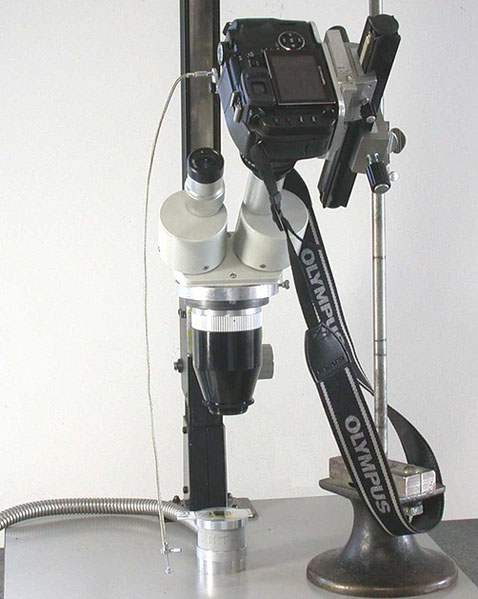
Photos 4 & 5 show the E-330 mounted with its 28 mm f/2.8 lens
almost touching the Meiji 10X eyepiece. My final alignment procedure
was to close the aperture diaphragm of the 28 mm camera lens until
the brightfield image in the camera, set for live viewing, became
seriously vignetted with a somewhat an out-of –focus image of
the aperture formed. Tilting of the bellows rail and rotating the
bellows rail on the ˝” diameter vertical shaft was used
to center the image of the aperture diaphragm and get its edge
uniformly imaged. The aperture was then fully opened. The 10X live
viewing from the main CMOS sensor was used for fine focusing of the
microscope. The spot metering does not work in manual mode with the
Olympus MF-1 adapter needed to mount the 28 mm lens. Live viewing
with the CCD sensor of the viewfinder system was used to obtain the
exposure readings in manual mode with no compensation with the camera
set for 100 ISO sensitivity. The test object chosen was the same
slide of mounted housefly wings used for my earlier article on using
the E-330 with my modified Biolam and 4X. The camera suggested
exposures were used to start a series of trial exposure to find the
optimum exposure, about 1/100 sec in brightfield and ˝ sec in
darkfield.
Figures
2 and 3
Figures 4 and 5
Photo 6 is a composite of the brightfield and darkfield images of the fly wings taken with a 0.5X reducer and the Meiji 2X objective. Some illumination unevenness is apparent towards the edge of the field. Photo 7 is a composite of the brightfield and darkfield images recorded using the Meiji 2X objective. Illumination is fairly even for this smaller field size. Photos 8 & 9 show the same wing in brightfield and darkfield recorded using the Meiji 4X objective. I am surprised to see the hair fringe resolved on the trailing edge of the wing was recorded. This fine detail is just resolved when viewing through the eyepieces. The tests indicated how well the critical focusing feature of the E-330 performs. The shutter vibration level is apparently minimal so that a very rigid stand is not needed for use with a stereo microscope. The photomicrography capability with the E-330 and my stereomicroscope is helpful but not necessary for me because both of my upright student microscopes have multimode illumination capability for 20X and 50X imaging with the E-330 mounted in my macro stand or on the enlarger stand. The modified Monolux microscope is described in my Microscopy Today article on rediscovery of darkfield dispersion staining. The dispersion staining image in that article of the entire field of Chrysotile asbestos on a reference slide was recorded with the drawtube mounted macro lens.
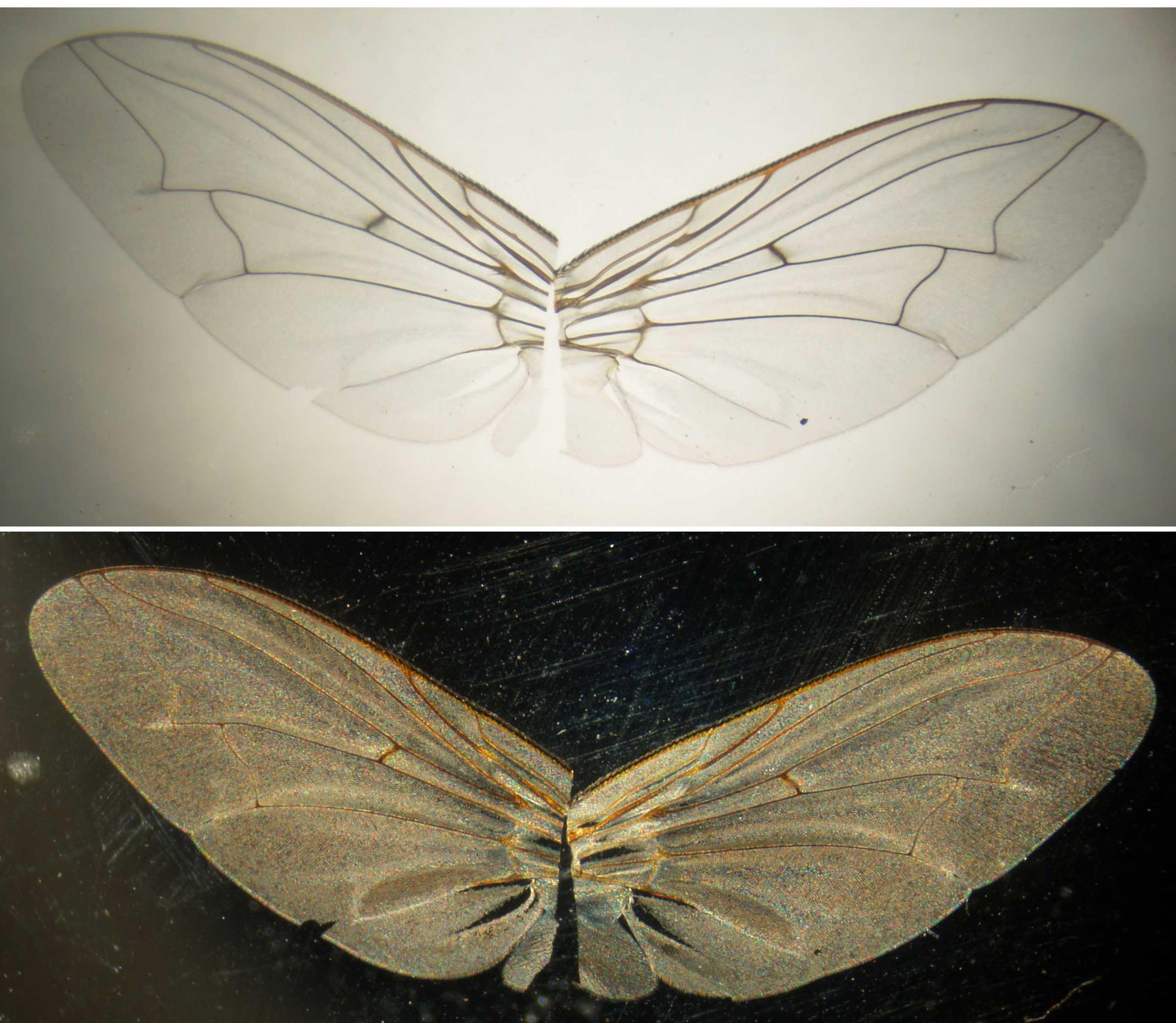
Figure
6 (Click image to view master)
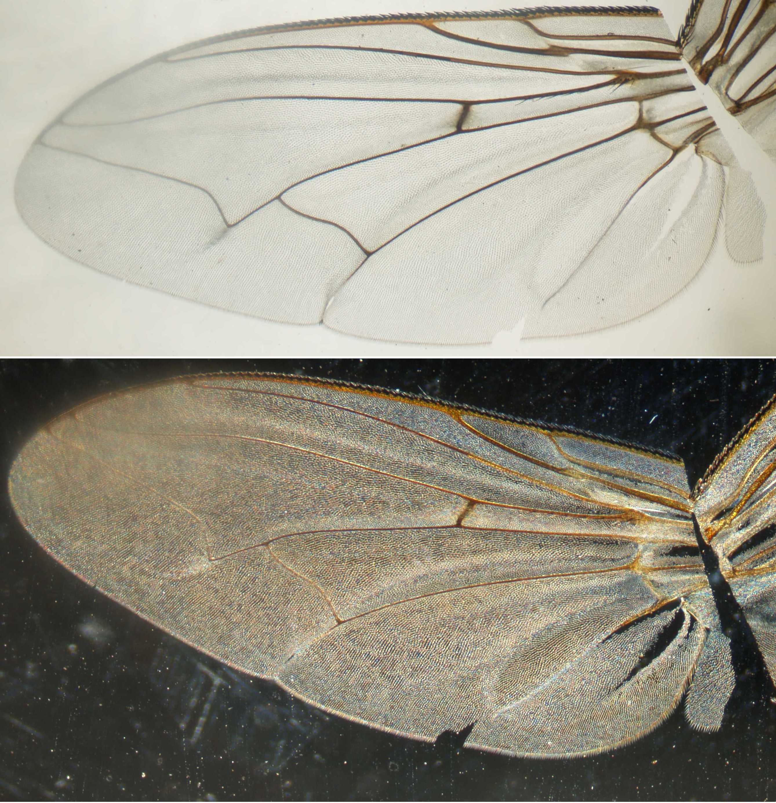
Figure 7 (Click image to view master)
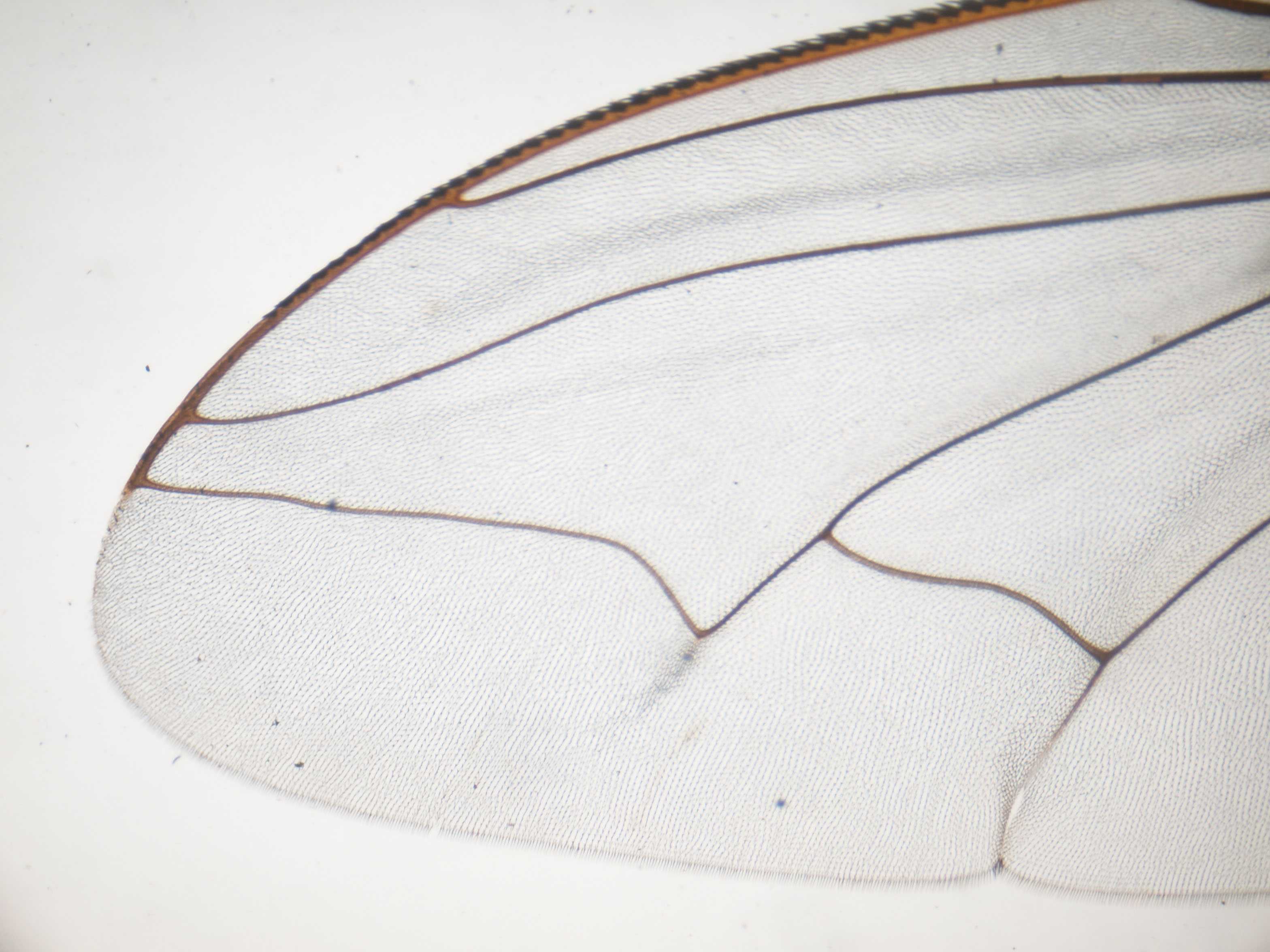
Figure 8 (Click image to view master)
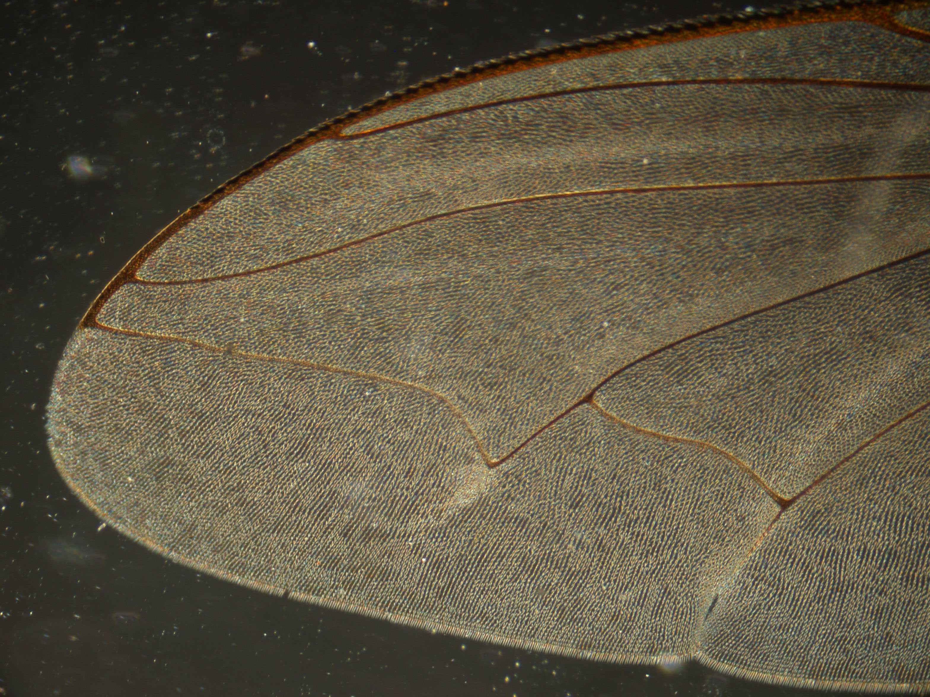
Figure 9 (Click image to view master)
Comments to the
author
Ted
Clarke
are welcomed.
Micscape Editor's acknowledgement: Local pdf copies of the author's 'Microscopy Today' articles are courtesy of the author and the 'Microscopy Today' Editor.
Published in the February 2007 edition of Micscape.
Please report any Web problems or offer general comments to the Micscape Editor .
Micscape is the on-line monthly magazine of the Microscopy UK web site at Microscopy-UK
© Onview.net Ltd, Microscopy-UK, and all contributors 1995
onwards. All rights reserved.
Main site is
at www.microscopy-uk.org.uk
with full mirror
at www.microscopy-uk.net
.