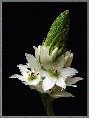
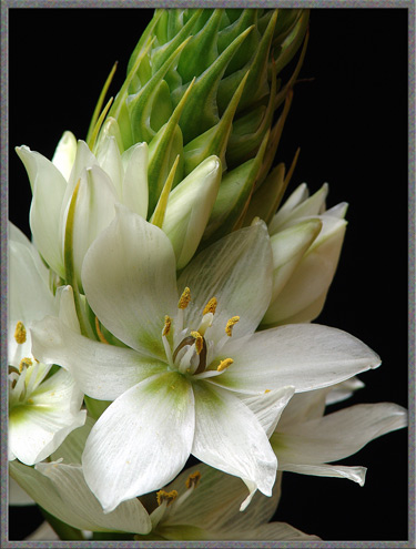
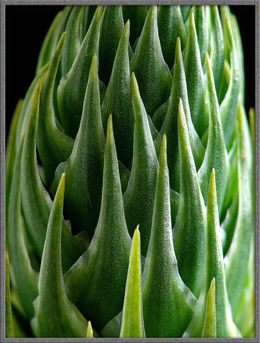
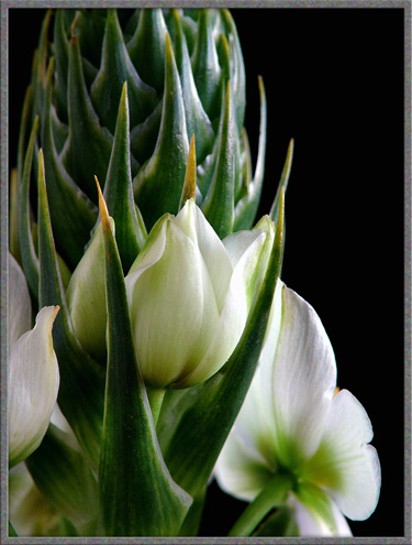
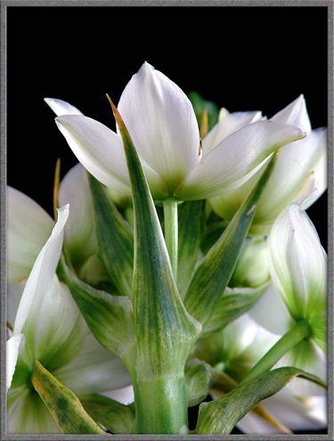
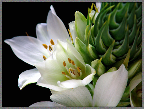
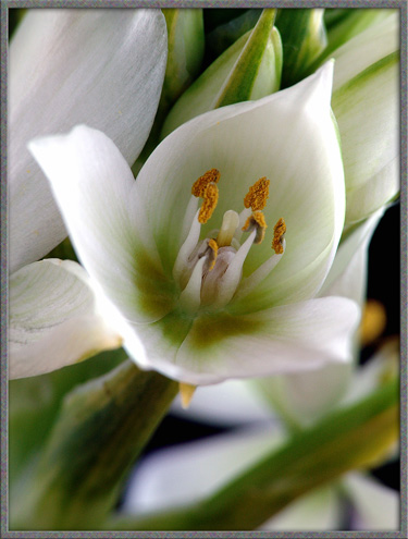
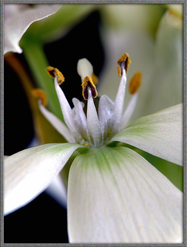
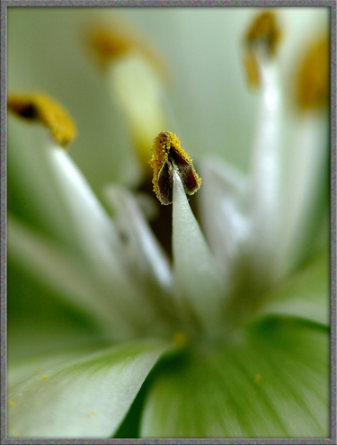
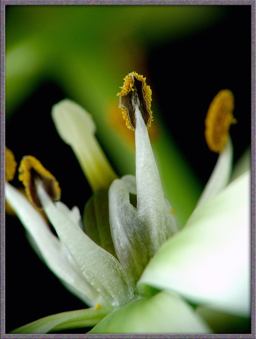
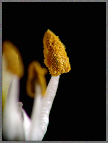
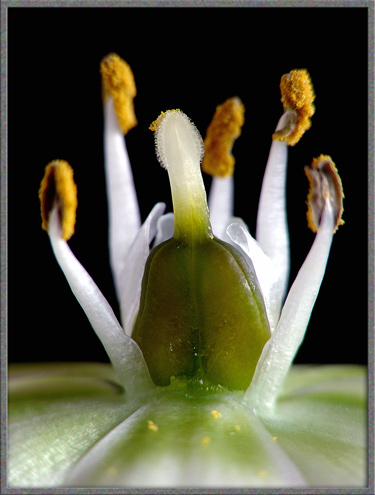
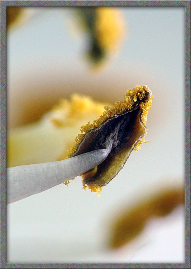
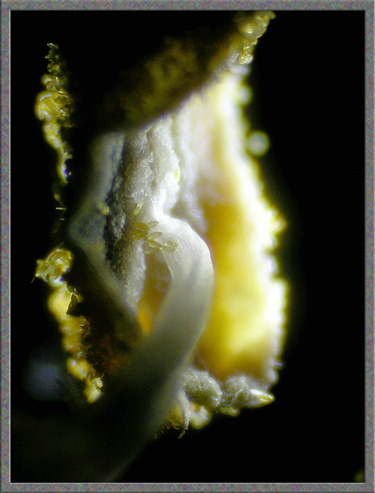
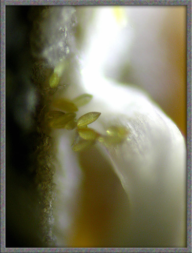
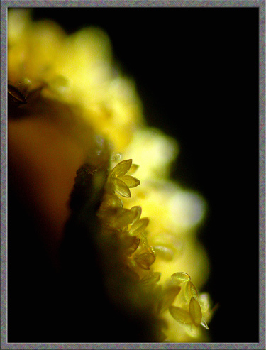
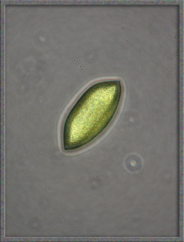
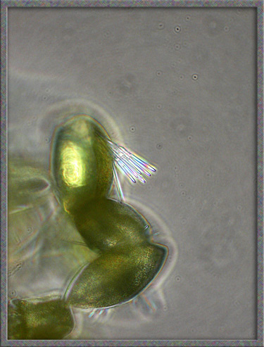
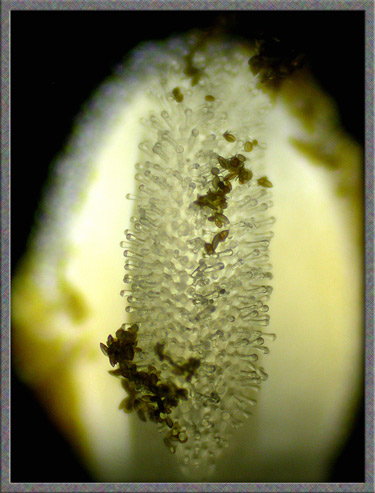
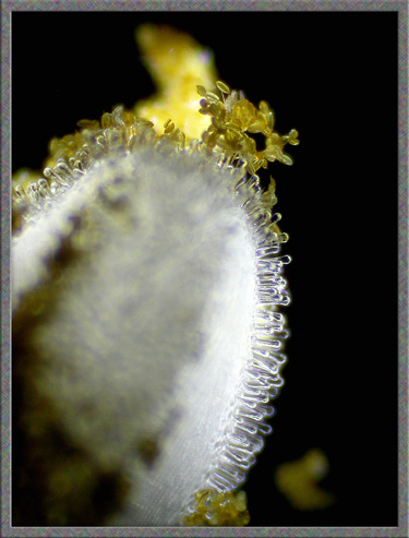
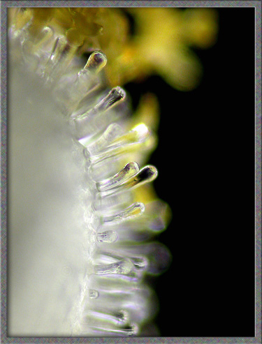
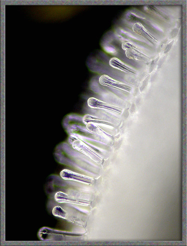
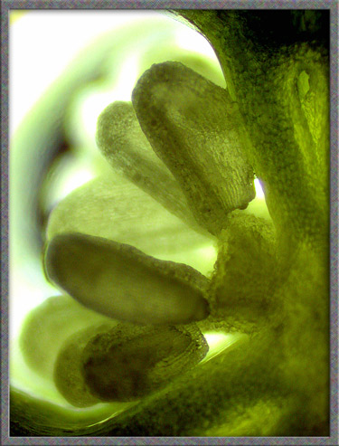
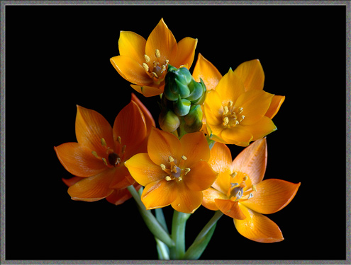
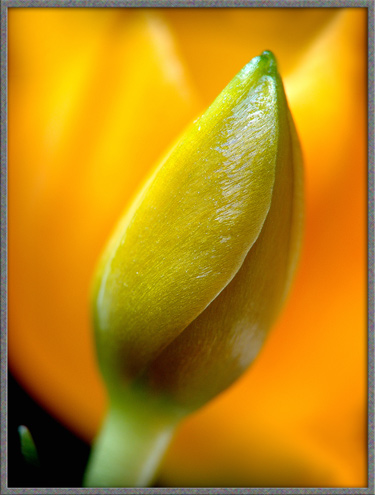
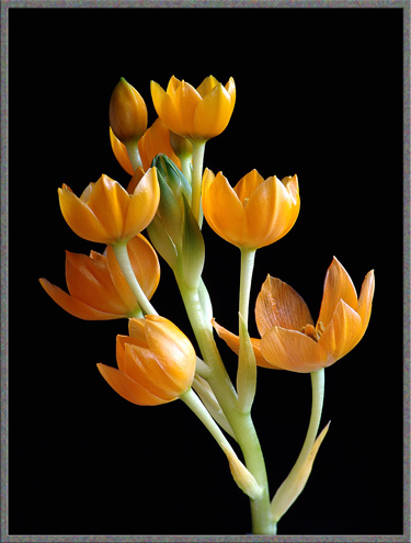
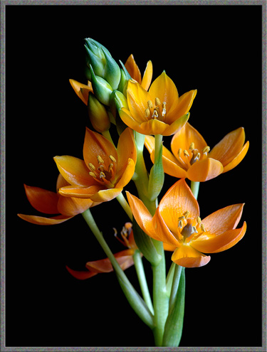
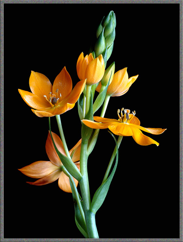
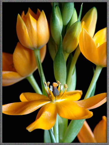
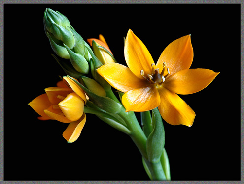
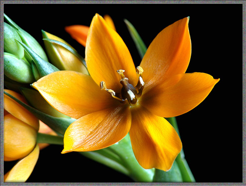
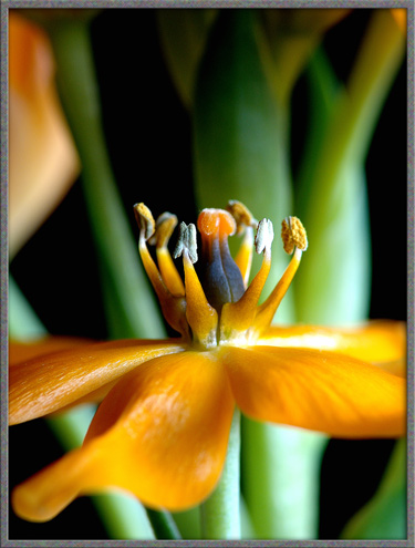
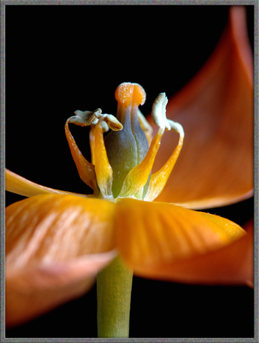
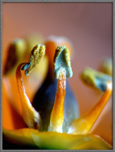
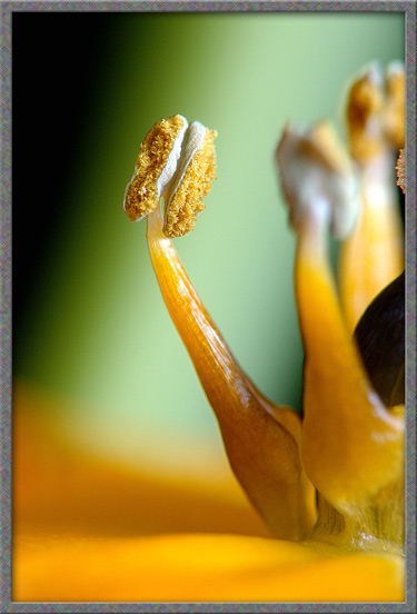
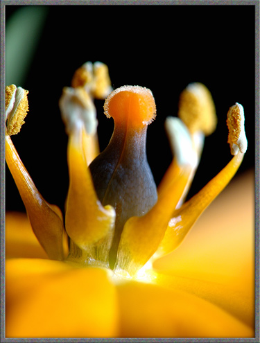
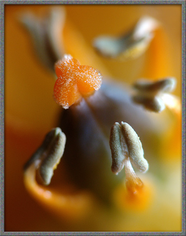
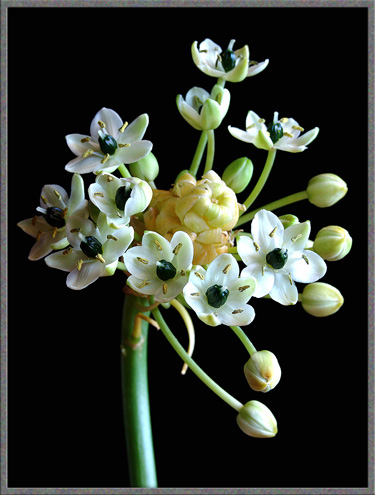
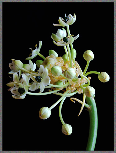
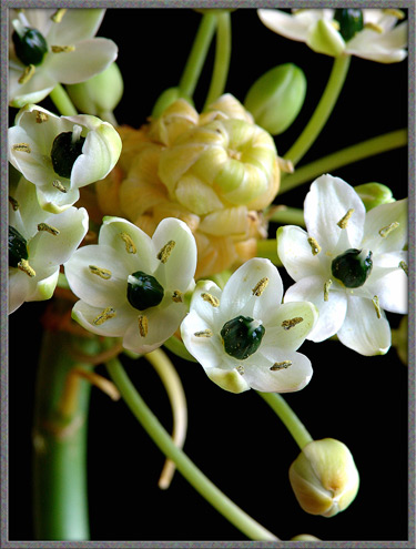
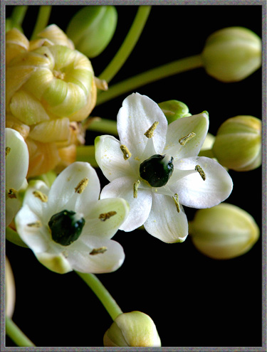
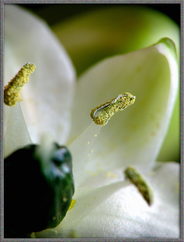
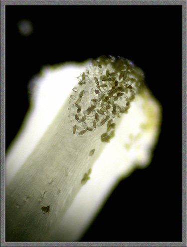
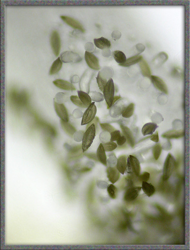
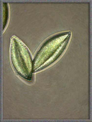
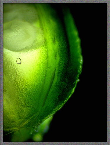
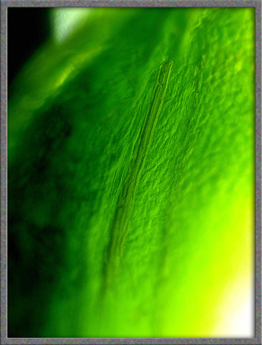
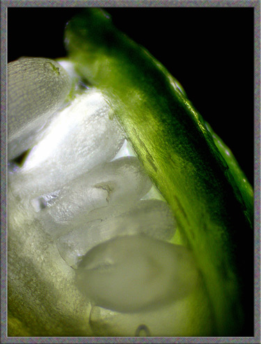
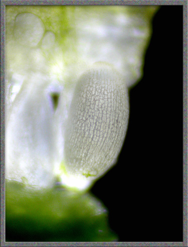
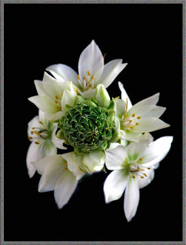
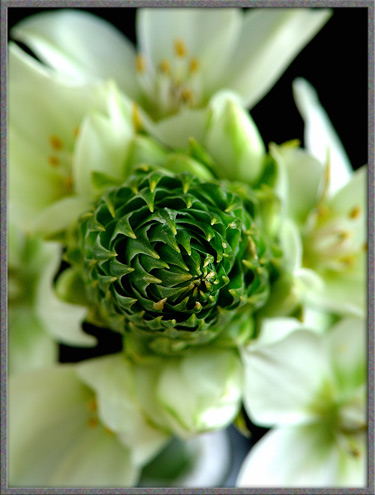
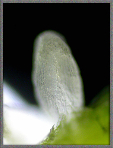
A Close-up View of Three Ornithogalum Flowers

"Chincherinchee", "Snake Flower" & "Arab's Eyes"

(Ornithogalum thyrsoides , Ornithogalum dubium
& Ornithogalum arabicum
)


by Brian Johnston   (Canada)


All three of the plants discussed in this article are cut-flowers purchased at a tiny “boutique” flower shop located in a large shopping mall in Toronto.  Florists sometimes refer to the three, tongue in cheek, as “florist’s nightmares”, since they often last for months when the cut stems are placed in water.  Although I prefer to photograph native wildflowers, Canadian winters prevent the activity from the middle of October until the beginning of May.  Out of desperation, I am therefore forced to photograph the wildflowers of warmer climates, in this case, South Africa and Western Australia.

The genus Ornithogalum previously belonged to the family Liliaceae.  It is now considered to be a member of the family Hyacinthaceae.  The three plants illustrated in the article are bulbous perennials, and are considered to be poisonous, as they contain Cholestane glycosides and calcium oxalate.

Chincherinchee [chin·che·rin·chee] – Ornithogalum thyrsoides

The Greeks referred to something incredible as being “bird’s milk”, ornithos – bird and gala – milk.  This seems to be the accepted derivation of the genus name Ornithogalum, and also one of the common names, “Wonder Flower”.  The extremely strange common name “Chincherinchee” comes from the fact that South Africans refer to the flowers as "tjenkenrientjee".  In addition to the previously mentioned common names, this plant is sometimes called the “Star of Bethlehem”.  (Unfortunately, many other unrelated plants are also called “Star of Bethlehem”, illustrating the danger of using common names alone.)

The first image in the article shows the typical pyramid-shaped cluster (raceme) of white flowers which grows on top of a leafless stem up to one metre in length.  (A raceme is an arrangement of stalked flowers.) The white blooms with pale green colouration at the centres, are composed of six petals.  As the flowers age, the green colouration tends to become lighter or disappears entirely.


The images below shows the very tight packing of the leaflets in the raceme, and the many encircling flowers.


The dark green raceme contrasts strikingly with the pale white flowers of the plant.  In my specimen, the raceme was about ten centimetres in length. 


Eventually, all of the tightly packed, green spear-shaped leaflets are pushed out by short-stalked buds.  The white flowers, which bloom from bottom to top, are large enough to completely obscure these green leaflets in the later stages of the blooming plant.  The raceme eventually displays up to about thirty flowers.


The short flower stalk, and faintly striped green bracts (modified leaves), can be seen below.


As a bud opens, the reproductive structures of the flower are revealed.  Each flower has six stamens consisting of an anther, (the male pollen producing organ), and a supporting filament.  A single pistil, consisting of a stigma, (the female pollen accepting organ), and its supporting style, is positioned at the centre of the ring of stamens.


Three images with increasing magnification, of the back of a stamen, are shown below.  Notice in the third photograph, the two translucent “wings” that are present at the base of the filament.


The front of each anther is completely coated with light brown pollen grains.


By removing one of the anthers, it is possible to obtain an unobstructed view of the pale white stigma (top), the yellowish green style (middle), and darker green ovary in which the seeds develop (bottom).


The reverse side of an anther, with the point of attachment to the filament, can be seen below.


Under the microscope, this point of attachment seems remarkably fragile.  Notice the pollen grains clinging to the anther’s surface in the image to the right.


When more highly magnified, (and using phase contrast illumination), the finely dimpled surface of the peanut-shaped pollen grain can be observed.


Many of the pollen grains in my specimen seemed to have “sprays” of needle-like crystals associated with them.  (I suspect that the crystals are calcium oxalate.)


The stigma has three-fold symmetry, with three longitudinal protrusions covered with thick, translucent hairs.  Pollen grains can be seen adhering to these hairs.


At a higher magnification, it is evident that each hair has a globular end.


A photomicrograph of a thin slice through the ovary reveals the developing seeds, (green oval structures).


An individual developing seed can be seen to be white in colour when it is removed from the surrounding green ovary covering.


When a stamen is pulled away from the flower, the connecting plant material tears away to form an extremely thin ribbon.  The microscope reveals the fine parallel strands that comprise the material.


Snake Flower
Ornithogalum dubium


This close relative of Chincherinchee possesses beautiful orange, cup-shaped flowers.  These flowers also have six petals, but the diameter tends to be smaller than those of Chincherinchee.

A single unopened bud can be seen below.


The cup-shape of each flower is evident in the left image below.  As the flowers age, they flatten out as in the right image.


Notice that the flower stalks in this species are longer than in those of Chincherinchee.


Although Snake Flower has a raceme, it is not as spectacular or as tightly packed as that of Cincherinchee.


Each flower has six stamens and a single pistil.


Notice that the pistil has a bright orange stigma, and that the style is so short that it is almost non-existent.  The ovary is a dark brown colour.


A higher magnification reveals the structure of a Snake Flower stamen.


The front of each anther is divided into two pollen covered lobes with white undersides.


Fine white hairs coat the surface of the stigma.  These hairs help to hold pollen to the surface after it has been transported to the flower by visiting insects.


The three lobed structure of the stigma, and two-lobed structure of an anther can be seen clearly below.


Arab’s Eyes – Ornithogalum arabicum


This plant has a very long leafless stem, (often more than a metre in length), with a roughly umbrella-shaped arrangement of small, waxy, white flowers on top.  The distinctive green-black ovary gives the plant its common name.  The flower stems are longer than those of the other two species, and the blooms are smaller in diameter.

A closer view shows the waxy flowers, and beige colour of the raceme of unopened buds.


The anther is, as usual, coated with fine pollen grains.


One of the three lobes of the stigma can be seen in focus in the image on the left below.  The image to the right is a more highly magnified photograph of the same lobe.


Phase-contrast illumination reveals that each pollen grain has a deep longitudinal groove on its surface.


If part of the surface of the ovary is peeled away, the developing seeds are just visible beneath a roughly textured translucent membrane.  (The small circle at the middle left of the image is an air bubble.)


The following photomicrograph shows surface detail on the ovary.


Developing seeds within the ovary can be seen below.


In Canada, the three species illustrated in this article are found only as cut-flowers.  Nevertheless, their long-lived, and strikingly beautiful blooms can provide interesting subjects for macro-photography during the long, cold winter!


Photographic Equipment

The photographs in the article were taken with an eight megapixel Sony CyberShot DSC-F 828 equipped with achromatic close-up lenses (Nikon 5T, 6T, Sony VCL-M3358, and shorter focal length achromat) used singly or in combination. The lenses screw into the 58 mm filter threads of the camera lens.  (These produce a magnification of from 0.5X to 10X for a 4x6 inch image.)  Still higher magnifications were obtained by using a macro coupler (which has two male threads) to attach a reversed 50 mm focal length f 1.4 Olympus SLR lens to the F 828.  (The magnification here is about 14X for a 4x6 inch image.) The photomicrographs were taken with a Leitz SM-Pol microscope (using a dark ground condenser), and the Coolpix 4500.  


All comments to the author Brian Johnston are welcomed.


Microscopy UK Front Page
Micscape Magazine
Article Library


© Microscopy UK or their contributors.

Published in the February 2006 edition of Micscape.
Please report any Web problems or offer general comments to the Micscape Editor.
Micscape is the on-line monthly magazine of the Microscopy UK web
site at Microscopy-UK  


© Onview.net Ltd, Microscopy-UK, and all contributors 1995 onwards. All rights reserved. Main site is at www.microscopy-uk.org.uk with full mirror at www.microscopy-uk.net .