|
|
A Close-up View of the Unusual Wildflower (Asclepias syriaca) |
|
|
A Close-up View of the Unusual Wildflower (Asclepias syriaca) |
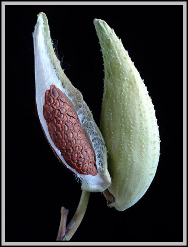
Most of us, I would surmise,
associate the term milkweed with the large, elongated tear-drop shaped
seed pods that the plant produces during late summer and early
autumn. This species however, is well worth observing during the
rest of its life cycle, for it appears remarkably different than its
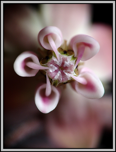
neighbours. Few wildflowers look like the one shown above!
The common name milkweed stems from
the fact that when cut or bruised, all parts of the plant exude a milky
white, sticky sap. This liquid contains poisonous glycosides
similar to those in Foxglove plants. When Monarch butterfly
caterpillars ingest this sap, they are not harmed, but their bodies
become bitter tasting and poisonous to predators. This protection
remains even after their transformation to beautifully coloured
butterflies.
Asclepias,
the genus name, derives from the name of the Greek god of medicine,
Asclepias, since milkweed was used historically in medical
formulations. Wart removal was one such application. The
species name syriaca refers
to Syria. At one time, it was hypothesized that milkweed
originated in that country. Today it is believed that the plant
was transported to Europe from North America.
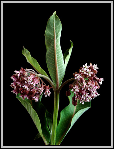
In early summer, buds begin to grow
on long drooping stalks that are connected to the same point on the
stem. Each cluster of stalks and buds is called an umbel. Notice in the image, that there are four umbels visible, each at a different stage of development.
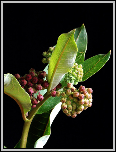
Young buds are light green, with just a hint of pink at the tip.
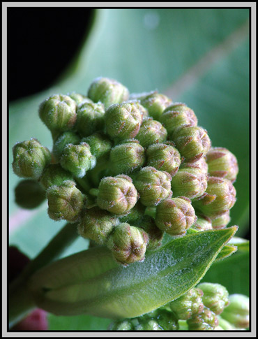
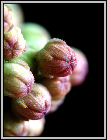
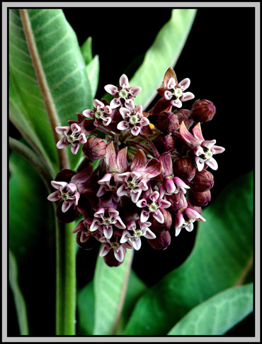
As they develop, the buds take on a
deep pink colour which becomes red just before blooming. (One
such umbel can be seen on the left of the second image above.)
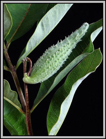
Milkweed leaves, which can be up to
ten inches long, grow in pairs and are positioned on opposite sides of
the stem. As can be seen in the close-up below, the leaves are
prominently veined. It is these leaves that provide the only food
for Monarch caterpillars. In fact, without milkweed foliage, it
is not possible for larvae to complete their life cycle.

In Ontario, milkweed flowers
bloom in July and August. Each globular cluster is 5 to 7 cm in
diameter and contains up to about thirty purplish flowers.
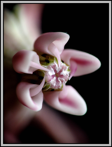
The flowers tend to be from 8 to 12 mm across.
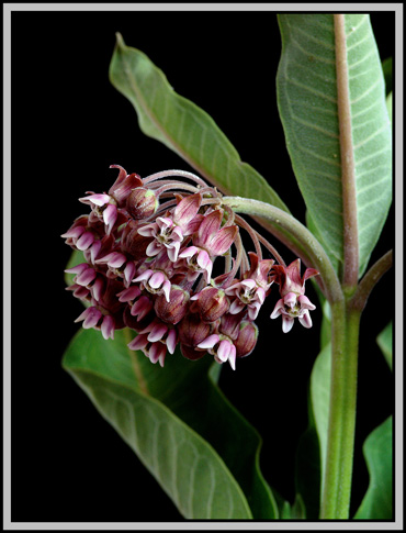
A closer view reveals the uniqueness of a milkweed bloom.

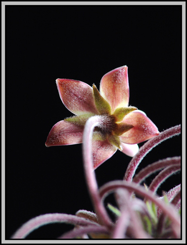
Notice in the left image below that
the flower stalks are very woolly. The image on the right shows
the five pale green sepals, or modified leaves, that bisect each pair
of petals.
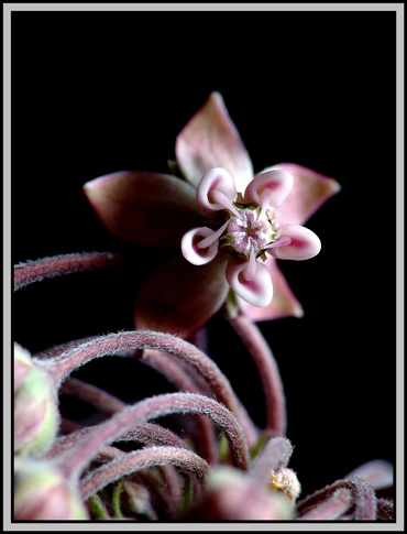
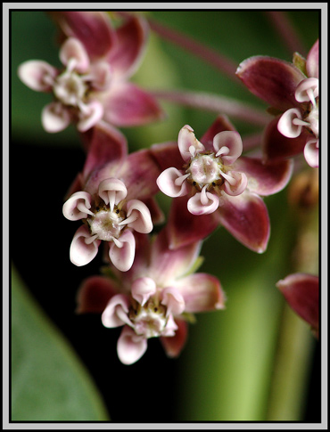
In a milkweed bloom, the five petals are bent backwards. The collective term for the petals of a flower is the corolla and each of the petals is called a lobe. In proper scientific terminology, it is the lobes of the corolla that are bent backwards!
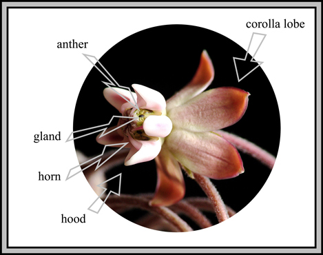
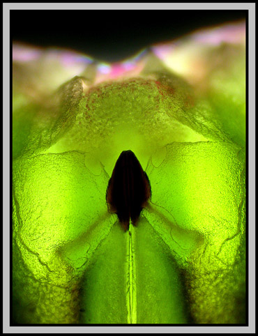
The corona,
which looks like a five lobed crown in the photograph below, is formed
by the fusing together of the five stamen filaments. Each lobe of
the corona consists of a tubular hood with a horn projecting from its
centre. Each of the hoods contacts its corresponding anther as
can be seen in the diagram.
Between each hood and anther combination, if you look carefully, there is a tiny dark elliptically shaped glandular structure.
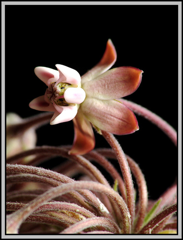
A photograph of the front of a
flower shows the five hoods and horns clearly. If you look
closely, a few of the dark glandular structures are just visible
between the hoods.


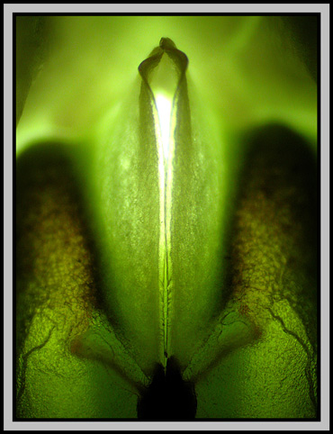
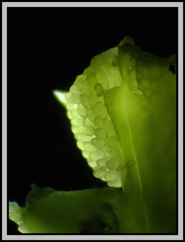
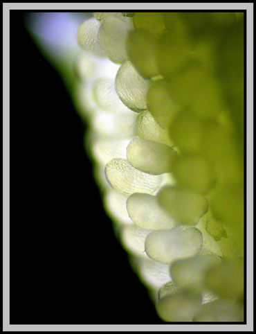
The two photomicrographs below show
that this gland forms the top of a slit-like opening between the
anthers. In the right hand image there are two orange brown
structures hidden behind the walls forming the slit. They are
joined to, and part of the gland.
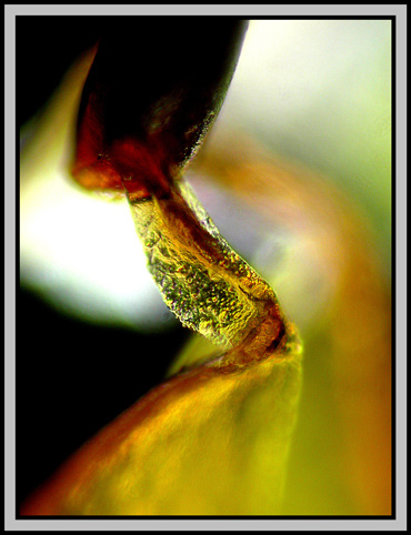
These sticky orange structures can
be seen in the image on the left below. On the right is a higher
magnification photomicrograph showing the point of attachment to the
elliptical external feature.
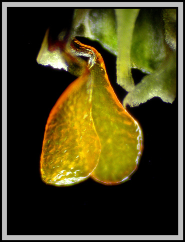
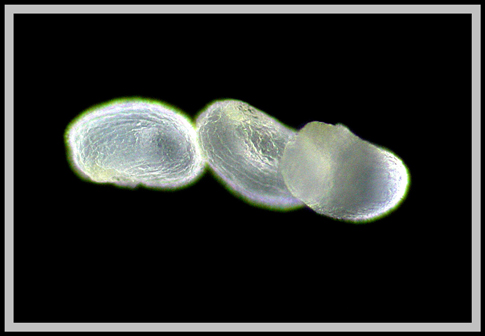
In milkweed, pollen is produced in waxy masses called pollinia
that dangle in pairs from the dark gland and are attached to the orange
structures seen above. Below you can see one of these
pollinia. On the right is a magnified section.
In the photomicrograph below, it is evident that the pollen grains are roughly elliptical and have a concave surface.
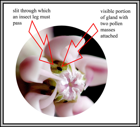
The pollination of a milkweed
flower is a very specialized procedure. (Use the image and
diagram below to visualize what must happen as you read the following
steps.)
Since this procedure is unusually
complex, very few of the flowers in each umbel produce fruit.
(This will be shown graphically later in the article.)
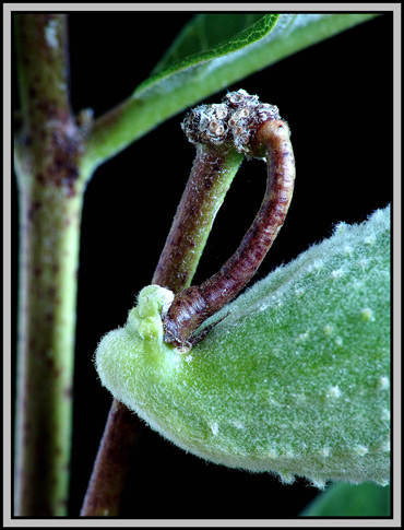
Milkweed flowers do not remain on
the plant for very long. After about a week or two, all of the
unpollinated flowers drop off. A long rough-textured green pod
called a follicle begins to
form at the location of the successfully pollinated flowers. The
stalk of each pod is strangely curled backwards as can be seen in the
image below.
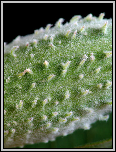
The surface of the approximately 12 cm long pod is covered in fine hairs and many soft protuberances.
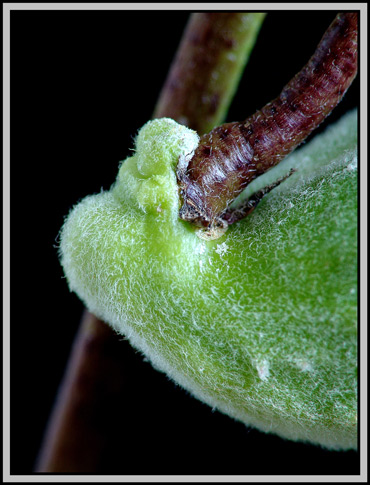
At the point of attachment to the stalk, the pod is strangely lumpy.
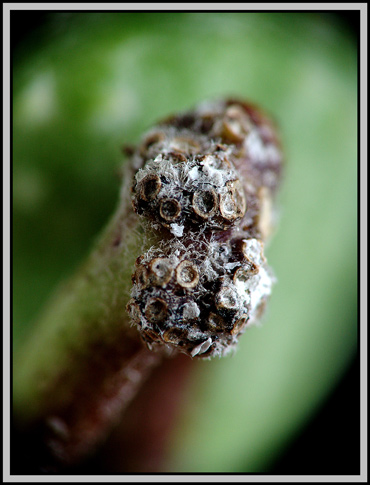
It is interesting to study the top
of the stalk holding the pod. One can clearly see the depressions
where the milkweed flowers were attached while blooming. Of the
approximately thirty flowers in this umbel, only one was successfully
pollinated! Clearly, chance does not favour insects as they
investigate the blooms.
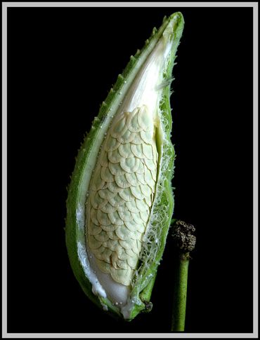
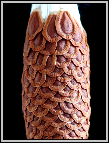
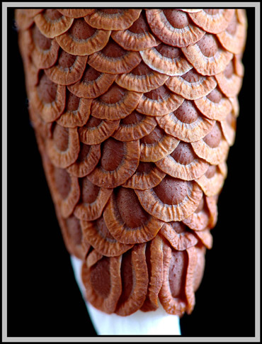
Within the pod, the approximately
150 overlapping seeds, each with its tuft of white hair, are
growing. The image on the right shows seeds with green centres at
a later stage of development. Note the thick walled shell of the
seed pod. The many fibres joining the two walls provide strong
but rubbery-feeling protection for the growing seeds.
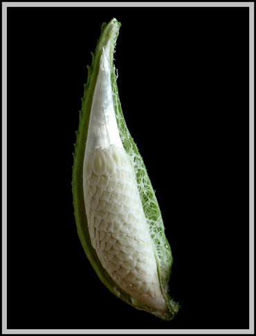
In the following plant, two flowers were successfully pollinated. At this point the seeds are almost mature.
The details of both top and bottom of the seed assembly are shown below.
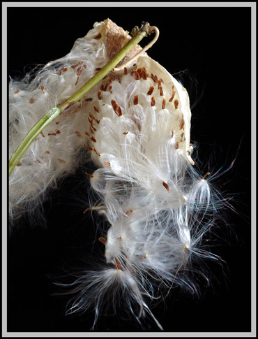
Each
pod eventually opens along a fold on one side. I was surprised at
how long it took for the pods to dry out and open on their own.
Patience was rewarded when after more than a month from the stage shown
above, the pods began spilling their seeds to the wind. The
leaves and stem had long since turned from green to yellow-brown at
this stage. In addition to propagation due to wind transport of
the seeds, milkweed also grows underground rhizomes that give rise to
new plants. For this reason, the plant frequently forms large
colonies.
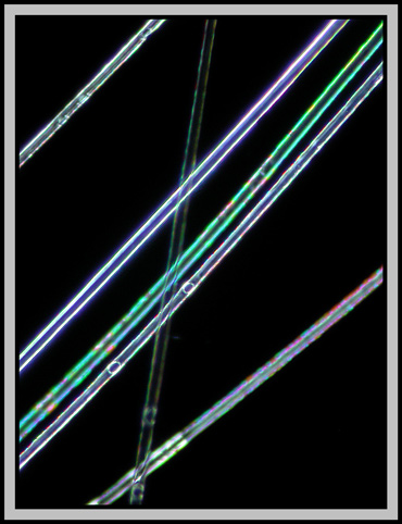
The image below uses a polarizing microscope with crossed polars to show some of the white hairs attached to the seed.
Milkweed is not a particularly
useful plant to humans, but try to imagine the world without colourful
Monarch butterflies. It is to be hoped that this plant long
remains an interesting part of the flora of our planet!
Photographic Equipment
The photographs in the article were
taken with an eight megapixel Sony CyberShot DSC-F 828 equipped with
achromatic close-up lenses (Nikon 5T, 6T, Sony VCL-M3358, and shorter
focal length achromat) used singly or in combination. The lenses screw
into the 58 mm filter threads of the camera lens. (These produce
a magnification of from 0.5X to 10X for a 4x6 inch image.) Still
higher magnifications were obtained by using a macro coupler (which has
two male threads) to attach a reversed
50 mm focal length f 1.4 Olympus SLR lens to the F 828. (The
magnification here is about 14X for a 4x6 inch image.) The
photomicrographs were taken with a Leitz SM-Pol microscope (using dark
ground and polarizing condensers), and the Coolpix 4500.
References
The following references have been
found to be valuable in the identification of wildflowers, and they are
also a good source of information about them.
Published in the February 2005
edition of Micscape.
Please report any
Web problems or offer general comments to the
Micscape
Editor.
Micscape is the on-line
monthly magazine of the Microscopy UK web
site at
Microscopy-UK
© Onview.net Ltd, Microscopy-UK, and all contributors 1995 onwards. All rights reserved. Main site is at www.microscopy-uk.org.uk with full mirror at www.microscopy-uk.net .