The images seen here show twenty species of foraminifera from Brian Darnton's collections from Terneuzen, on the Westerschelde River estuary. They have been selected to illustrate the remarkable variety of forms present at the site. Brian Darnton's corresponding article in this month's edition of Micscape gives a detailed account of the site; click on the link below for a description.
The Microscopic Virtues of Mud.
The drawings have been rendered using scraperboard. Scraperboard is a medium rarely used for scientific illustration, but one that I felt was particularly appropriate; since foraminifera tests are white or light in colour, they contrast well with the ground of black scraperboard. The medium, which produces an effect reminiscent of engraving, also allowed for the execution of fine detail such as the many perforations in some of the tests, and the representation of a wide range of textures and differing levels of transparency, opacity and lustre.
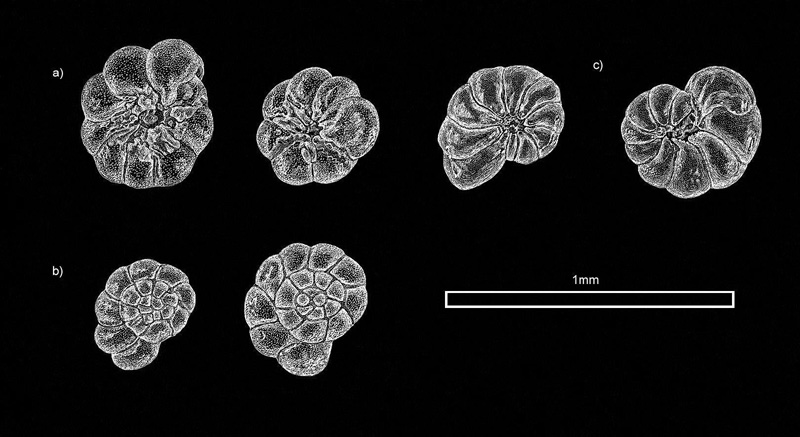
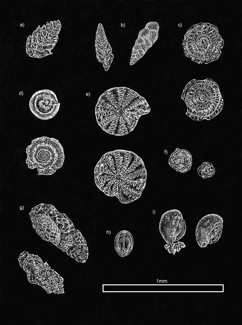
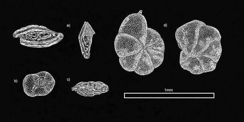
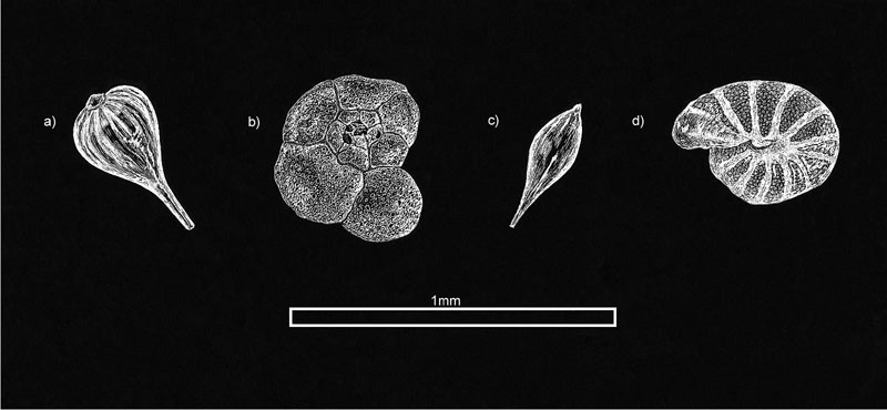
A scale bar equivalent to 1mm is included in each illustration. The foraminifera appear in, and have been grouped according to, descending order of the frequency with which they occur at the site.
Dominant species, making up more than 25% of samples.
a) Ammonia beccarii Linne (umbilical); b) Ammonia beccarii Linne (spiral); c) Protelphidium germanicum Ehrenberg. The above are typical estuarine species.
Species present in most samples.
a) Brizalina spathulata Williamson; b) Brizalina variabilis Williamson; c) Patellina corrugata Williamson; d) Spirillina vivipara Ehrenberg; e) Elphidium willansoni Haynes; f) Gravelinopsis praegeri Herron-Allen; g) Eggerella scabra Williamson (note the agglutinated test made up of cemented, mainly uniformly sized grains); h) Fissurina lucida Williamson; i) Milionella subrotunda Montagu.
Species rare but present.
a) Spirosigmolina pusilla Earland; b) Rosalina globularis d'Orbigny; c) Trifarina angulosa Williamson; d) Cibicides lobulatus Walker & Jacob. N. B. Attached to the upper half of the smaller Spirosigmolina foraminiferan is part of a frustule of the diatom Triceratium favus!
Single example only found.
a) Lagena perlucida Montagu; b) Trochammina inflata Montagu; c) Lagena clavata d'Orbigny; d) Melonis pompilioides Fichtel & Moll.
Species identification by Brian Darnton.
A Web page that provides a good basic introduction to foraminifera can be found at UCL's website at the following address: http://www.ucl.ac.uk/GeolSci/micropal/foram.html. This resource covers classification and basic descriptive terminology, the occurrence of foraminifera in the fossil record, and their use in stratigraphy and other environmental disciplines. Also displayed are photomicrographs of a selection of planktic and benthic foraminifera, taken using both SEMs and the light microscope.
Christina Brodie is currently writing and illustrating a book about botanical illustration techniques, Drawing and Painting Plants, to be published by A & C Black (London) in August 2005.
Please contact the author, Christina
Brodie via her website www.queen-christina.com if there are requests about image use or if interested in
her other artwork.


