Bougainvilleas spectacular floral
display is admired throughout the world. Native to tropical South
America, Latin America, the Caribbean, and Southeast Asia, it can grow
in a small pot, be a sizable tree, or spread like a vine over a
wall. In cooler climates it grows happily as a house plant, or
hanging basket. It certainly is versatile!
Bougainvillea was discovered in the
1760s in Rio de Janeiro by a French naturalist named Dr. Philibert
Commercon. He named it after his friend Louis-Antoine de
Bougainville, a ships admiral and the captain of the vessel that
carried him around the world in a voyage during the period 1766 to
1769. In the early 19th century Kew Gardens in England
played an important role in disseminating Bougainvilleas that it had
propagated to British colonies throughout the world.
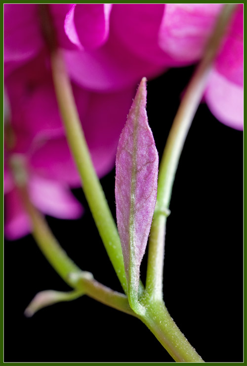
The particular hybrid studied in
this article, Bougainvillea glabra
Vera, has brilliant red blooms and a compact habit. It
can be seen in the image above. Additional images follow that
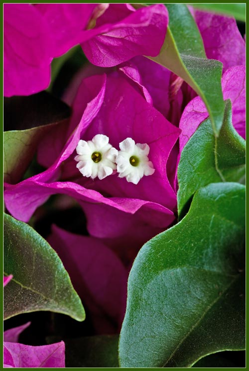
show closer views of the colourful flowers. Well, not
really. Only a couple of the images show Bougainvillea
flowers! The brilliant red structures are not the flowers petals
at all, but simply the protective bracts (modified leaves) that
surround and protect the real flowers! The real flowers are the
insignificant tubular, cream-coloured structures visible in a few of
the images. (Another similar situation exists in the Poinsettia
plant.)


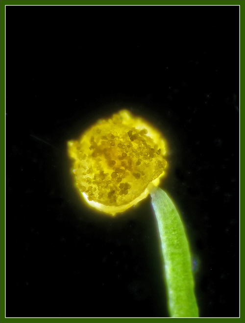
I
Photomicrographs showing these
protuberances can be seen below. As usual, the protuberances
increase the stigmas surface area and help it to acquire and retain
pollen grains.
If a scalpel is used to remove the
outermost layer of tissue from the corolla tube, and the tissue is
examined under the microscope, the view is as follows.
If a vein on the base of one of the
bracts is examined microscopically, the tiny red hairs that can be seen
below are visible.
It is estimated that there are over
300 varieties of Bougainvillea worldwide, in colours as diverse as
white, pink, orange, purple, burgundy and of course, bright red.
Its unique and graceful form is certainly a super-star in the botanical
world.
Photographic Equipment
The low magnification, (to 1:1),
macro-photographs were taken using a 13 megapixel Canon 5D full frame
DSLR, using a Canon EF 180 mm 1:3.5 L Macro lens.
An 10 megapixel Canon 40D DSLR,
equipped with a specialized high magnification (1x to 5x) Canon macro
lens, the MP-E 65 mm 1:2.8, was used to take the remainder of the
images.
The photomicrographs were taken
using a Leitz SM-Pol microscope (using a dark ground condenser), and
the Coolpix 4500.
Update June 2023. Sadly the author passed away a few years ago. A valuable comment is added below.
Beautiful images. Comment: The flower tube in which the stamens and pistil are found is actually the calyx, not the corolla. These plants have no corollas. The twisting of the tube after flowering is not a genetic defect, but is found naturally in several species of Bougainvillea. I suspect it is to close off the calyx tube to protect the developing ovary. Michael H. Nee, PhD
A Flower Garden of
Macroscopic Delights
A complete graphical index of all
of my flower articles can be found here.
The Colourful World of
Chemical Crystals
A complete graphical index of all
of my crystal articles can be found here.