MICROSCOPES, FILTERS, DIAPHRAGMS AND STOPS
Technical details for Micscape article HOMAGE to the ONION SKIN
with some additions
Walter Dioni, Mexico
|
|
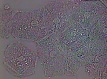
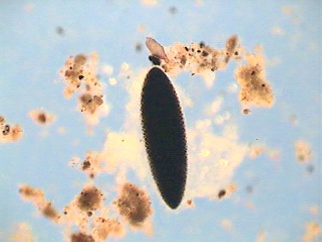
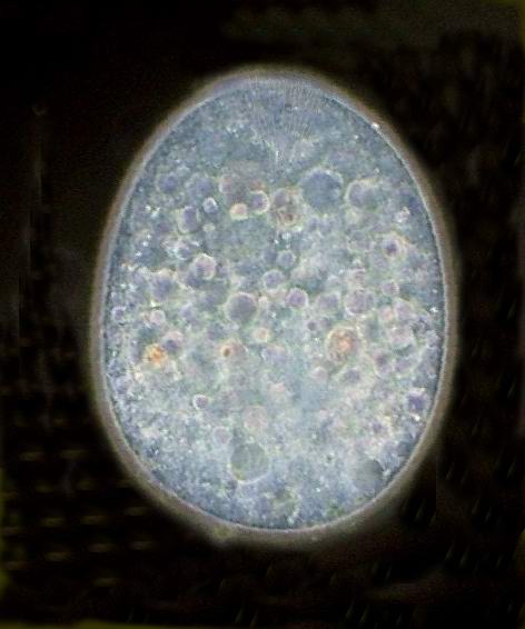
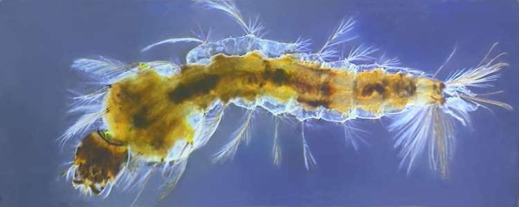
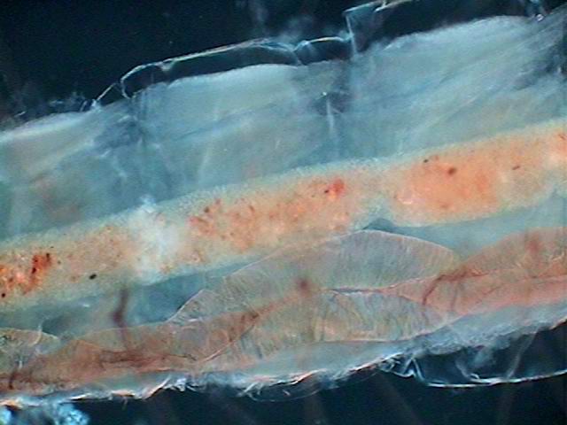
| Anopheles mosquito larvae, malaria parasite vector. 4x Objective. 3 pictures stitched with PhotoPaint.- COL-D |
EQUIPMENT- All my images have
been taken with a National Optical DC3-163P microscope, with an
integrated DC·3 digital camera, with automatic white balance and
controlled by its own capture software. The microscope is equipped with
DIN planachromatics 4x (NA 0.10), 10x (0.25), 40x (0.65) and 100x HI
(1.25) objectives, an Abbé condenser NA 1.25 and one 12V, 20W
lamp included in the base and capable of critical illumination. More
details on the instrument, the camera, and the software, can be
consulted on this site. www.microscopeworld.com
PHOTOGRAPHS. - The original format of capture is 640 X 480 px but have been resized in this article. For those who wish to take measurements on my pictures I tabulate below the width in micrometers (horizontal dimension) of the field of view of the microscope covered by the picture with each objective.
|
objective |
microns |
|
X4 |
3400 |
|
X10 |
1333 |
|
X40 |
340 |
|
X100 |
133 |
If you print the pictures, it is very easy to obtain an index of microns per millimetre,
by dividing the microns corresponding to the objective by the width in millimetres
the printed photo has.
ILLUMINATION
Bright field.- I have provided my illumination system with a permanent field diaphragm (FD) for the 4x objective, located over the front lens of the lamp and provided with the supplied blue filter (I made two interchangeable mountings to also use the green filter, I have also a yellow filter, but I never use it). It is very amusing, but Hellman's Mayonnaise 200g bottles have lids which fit exactly over the external ring of the front lens of my illuminator, so that they were only waiting for me to create the 30 mm opening which I need to adjust the blue or green filter in its interior. I adjust the filter with two rings of paperboard. That puts the surface of the "filter lid 3 mm over the front lens. If you don't like my solution you can find many more alternatives in this article by Rosemarie Arbur.
And I also made an individual set of field diaphragms (FD), according to
Ian Walker's Topical Tip.
 |
|
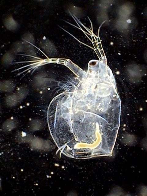
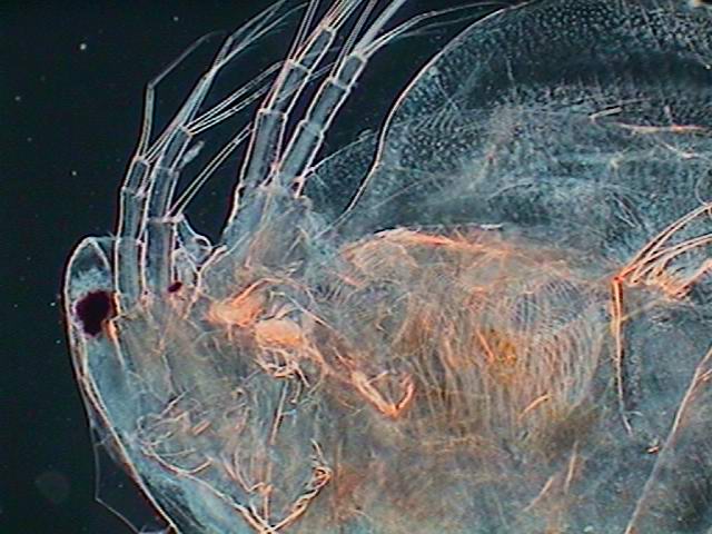
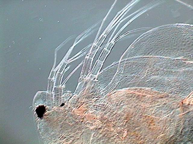
| Daphnia - x10 objective |
Cheek
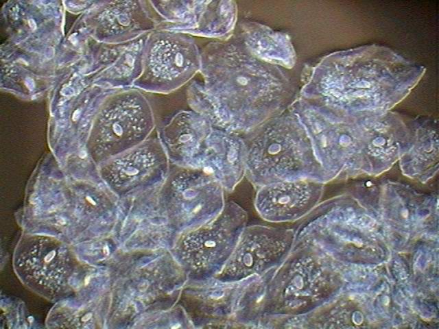
epithelium cells - 100x obj.- cropped from the 640 x 480 original -
contrast raised and lowered intensity for better displaying the cells. |
I reduced the number of FD's to only two: one of 13 mm for the 10x and other of 6 mm for the 40x and also for the 100x. A smaller diaphragm causes in my system undesirable marginal diffraction. I focus on a slide. Then I place on the blue filter one transparent acetate disc with a central cross and moving the condenser I focus this cross on the subject plane, then I change it for the
FD which corresponds. And I adjust the aperture diaphragm to
75% of the numerical aperture. I marked in white the positions of the
iris lever on the underside of the condenser, and mounted on the foot
of the microscope (which has a very suitable surface) a very small
mirror to see the position of the lever without undertaking acrobatics.
 |
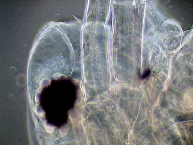
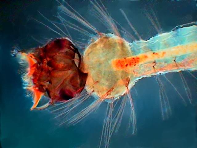 |
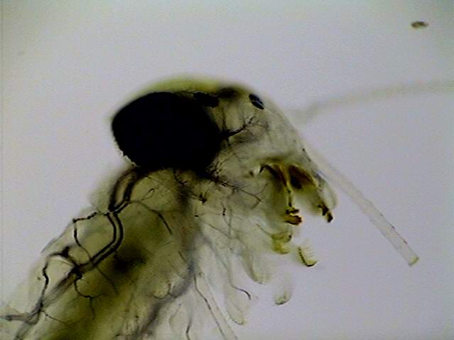
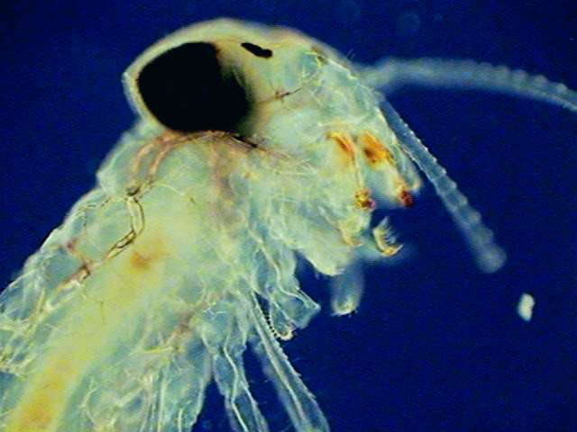
| Mosquito larvae - 4x objective |
Mosquito larvae - 4x objective- COL-D1 |
This
technique focuses on the preparation a disc of pure light, without
texture or dust. It is a modern version of the critical illumination,
of course.
One cannot make Köhler illumination without having an adjustable front lens
on the lamp, to focus the filament of the lamp in the field of the diaphragm of aperture.
If you have one, you can mount on the front lens of your illuminator an iris diaphragm, which is more convenient than the individual diaphragms.
FILTERS, DIAPHRAGMS AND STOPS
These are tools used in the condenser filter tray
One filter is a transparent disc, clear or coloured, interposed in the beam of light that enters the objective.
One stop is a disc with one or more opaque portions to interfere with selected portions of the same beam.
One diaphragm is an opaque disc or annulus with a transparent disc (central or eccentric). The opaque annulus can be fixed, or of continuously variable size (as for the iris diaphragm).
They are used to vary the form in which the light shows the object in the field of vision of the microscope, by giving contrast or colour to it. These discs are of the diameter of the filter tray (approx. 30 mm).
When you come to use these items you generally must forget the rules of Köhler illumination or Critical Illumination. Each one of them has its own rules of use to obtain the desired effect: an optimal height of the condenser, full or almost full aperture of the condenser diaphragm, and a suitable light intensity. And surely to be able to record the effect of the device in the illumination of your object you must also finely regulate the capture parameters of your software, especially the brightness and contrast, and also the colour balance many times.
You must remember that the filters, the diaphragms and the stops behave very differently depending on the subject. Its nature, thickness, texture, whether alive or fixed, if it is in the air or in a mounting medium, are determining factors for the effect of the tool used. What gives outstanding colours with a thick subject, can give a grey image with a very fine subject. A diaphragm which gives optimal results with some subjects may not be suitable for another. And a golden rule is that one must always start with the simpler black field stops. Many times that is enough.
| This is not a science, it is an art. The only guarantee of success lies in experimentation. But these tools are those which drives the amateur microscope with a power very close to that of much more expensive professional microscopes. If you do not have too much money you need these small tools, which give such fine and beautiful images. |
BLACK BACKGROUND I obtain the black background with black field stops (BFS) made as many authors have described. My BFS were
made by printing them on acetate with a HP ink jet printer with 1200
PPI. To obtain the adequate density I cement two or three copies with
nail varnish, applied where it does not obstruct the utility of the
stop. That also gives them more rigidity. My equipment includes a
series of 10 mm, 12.5 mm, 15 mm and an occasional 20 mm. The latter is the only BFS which gives me a real dark field with the x40 objective.
Until I used this big stop I believed that my 40x could not give a true
dark field. But as I mentioned above one must choose carefully the
preparations which can succeed.
 |
|
Daphnia, alive, 4x, 12.5 DFS |
You must regulate very finely the brightness, with the camera software, to record a true black background and to give to the subject the colour and detail which it shows under the microscope. It may be that you must decrease the contrast to capture the true texture of the objects. In general the 4x supports only dark field. But with some subjects, the 10x working with the 10 mm BFS can also provide a kind of marginal oblique illumination (see below).
Moreover, all DFS slightly moved off its centred
position can give sometimes very effective oblique illumination
effects, but surely with uneven illumination which can lower the
display quality of the image. Many times one can treat the background
in an image processor to eliminate the gradients.
 |
 |
| Daphnia, 4x obj. 10 mm centred DFS |
The same, 15 mm DFS off centre |
Micscape References (Dark field)
F.A.S. Sterrenburg
www.microscopy-uk.org.uk/primer/index.htm
Mike Samworth and Wim van Egmond
www.microscopy-uk.org.uk/mag/art97b/wimdfield.html
MARGINAL OBLIQUE ILLUMINATION.- Some dark field central stops are used with the 40x and, on some occasions, and with some subjects, with the 100x, to obtain the Marginal Oblique Illumination (MOI) which allows the effect of pseudo phases contrast described by Sterrenburg, and which is also the basis of Circular Oblique Illumination (COL) described by Paul James. To obtain the MOI one must put the condenser, with the filter well centred, at a height which makes it possible to block almost 95% of N.A. of the objective. With the field and aperture diaphragms completely opened one obtains the phase-contrast-on-the-house according to Sterrenburg, or at least a very good oblique illumination. In these cases the background is not black, but it is very pleasant.
F.Sterrenburg (phase contrast on the house)
 |
|
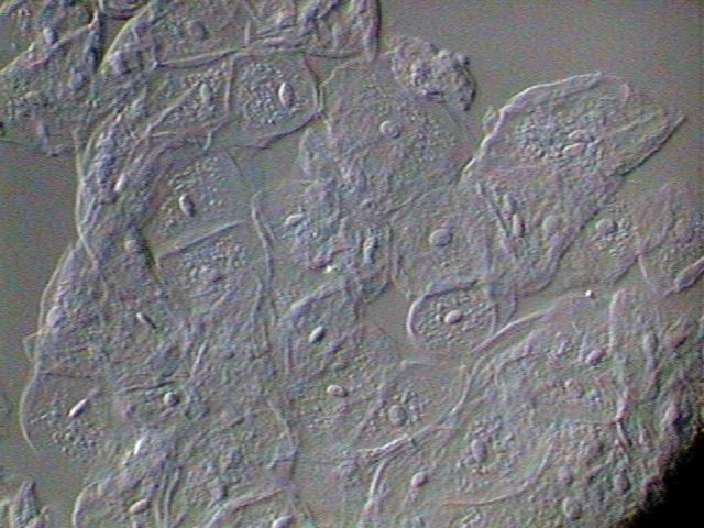
Cheek cells, 100x obj, - MOI |
Poor man's DIC - If used carefully, some DFS used according to these techniques produce with many material, results which resemble the images of DIC
(but not to be confused with the artificial results of some image processing software which also
produces the illusion of relief from flat images.)
But whatever the ObI or DFS you use, never let go the opportunity of slightly offsetting them with small displacements of the filter tray: one always obtains very interesting nuances.
For the young
new microscopists there is an interesting, cheap and easy to make
technical alternative (Mathias' Wedge) whose effects were discussed in
another article.
|
|
 |
| Daphnia head, 40x obj. - ObI - 10 mm marginal clear disk on 30.5 opaque stop. |
Cheek cells, 100x obj. half reduced Mathias wedge. |
References (Oblique illumination)
Paul James
www.microscopy-uk.org.uk/mag/artfeb00/pjoblic.html
Walter Dioni
RHEINBERG.- Rheinberg filters (RhF) have been prepared by the same method on acetate. One must remember that the central disc, that can be black or coloured, determines the background colour of the microscopic image, and that the peripheral ring obliquely illuminates the objects and gives them their colour. For better success the central disc must have the diameter of the DFS that corresponds with the objective in use. The density of the colours must be relatively intense, especially that of the central disc. One must make Rheinberg filters by superimposing and gluing two or three copies of the same disc or certain additional ones. For example one can glue two filters that have blue centres and red contour, plus one clear with blue centre to finish with a disc which has the centre denser than the periphery. One can easily make combinations of these transparent filters to obtain other effects.
On the use of Rheinberg filters one can consult
Chuck Huck
www.microscopy-uk.org.uk/mag/artsep00/chillumin.html
Wim van Egmond
www.microscopy-uk.org.uk/mag/artapr02/contrast.html
I do not agree with his methods to make the filters, but they give a good explanation of their use.
When the subject and the filter are well matched the results of RhF are spectacular and can be improved only by the COL diaphragms. But generally RhF
are effective with the 4x to 20x objectives. One can use them with the
40x, of course, but it is more difficult to appreciate the effects.
|
|
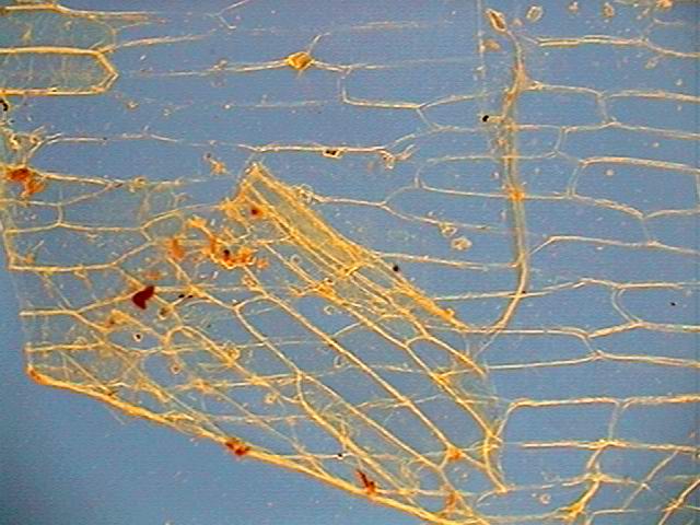
|
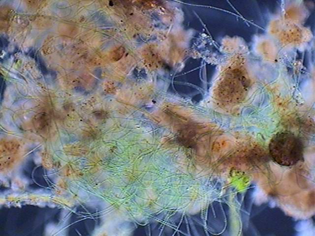
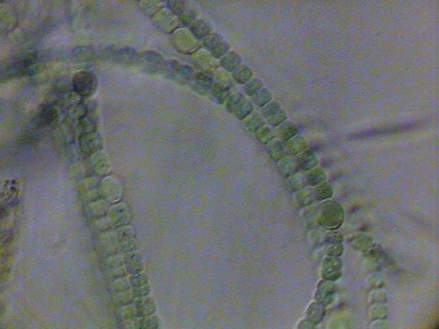
| Onion skin - 10x obj.- RhF - blue center - red annulus |
Anabaena - 10x obj. - RhF - blue center - clear annulus |
|
|
| Anopheles egg - 10x obj. - blue center -yellow annulus |
CIRCULAR OBLIQUE ILLUMINATION.- Paul James described sufficiently and thoroughly the use of stops for Circular Oblique Lighting (COL)
to obtain very interesting effects of illumination with the high powers
of the microscope. He does not discourage their use with the 10x and
20x objectives, but centres his attention on its effect with the 40x
and the 100x.
|
|
|
| Larvae of Ephemeroptera -4x obj. brightfield |
The same - COL -J |
|
|
| The same -COL-C |
His diatom pictures with COL diaphragms are exceptional (the stops he proposes is here called COL-J). The COL-J
which I generally use has a black central disc: 12 mm, followed by a
clear circle of 3 mm, a black one of 2 mm, another light of 2 mm and
black outside. It is a really effective stop, not only for the high
powers as Paul James suggests, but for several subjects with the 4x and
the 10x also.
 |
| Algae (Zygnema and Spyrogira) - 40 x obj. - COL-J |
There is also a COL-C,
because Christian Colin used a slightly different and simplified
version, (central disc of 12 mm, clear ring of 3 mm, black of 4 mm and
the external one clear), and some COL-D, because I made some modification to the COL stops and generalized its application to all the powers from the 4x to the 100x.
In most
subjects the COL-C give very similar results with COl-J. The difference
is something like the one betwen a sweet and a dry wine.
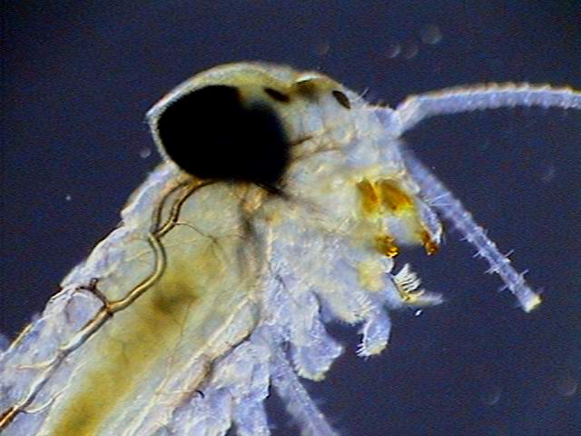
 |
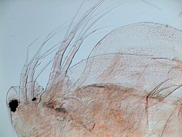
|
Detail, mosquito larvae - 10 x obj - COL-C |
The COL-D's are polychrome COL stops. I use stops that could have even the centre coloured and also the concentric rings. Blue is the more useful colour, and the stops which I more frequently use are: one with a central black disc of 11 mm, a clear ring of 3 mm, the following one blue of 4 mm and the peripheral ring clear; and the other with also the central disc black, but of 15 mm, and a clear ring of 3 mm, one blue ring of 2 mm and the following clear.
A disc which gives with some subjects extraordinary results, very similar to some phase contrast images, (it makes it possible to differentiate with very good definition the bacteria on the surface of organisms or cells), is a disc like the first described, but with a yellow or orange background.
There is an idiosyncrasy. It gives good results in direct vision, but my digital camera does not register faithfully the colours it gives. The sensor seems to be excessively sensitive to the red or yellow tints and the background is often mottled with yellow.
A very interesting COL-D is a COL-J, but with the interior clear ring coloured red, and the outside one blue.
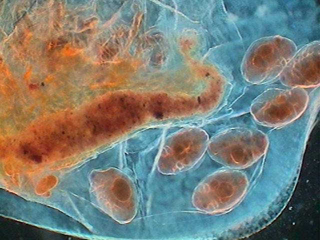 |
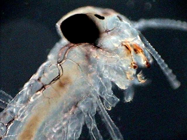
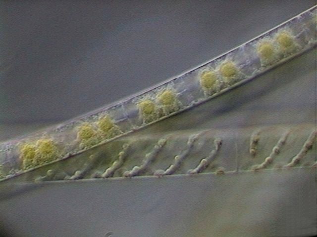
 |
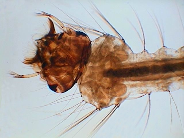
| Daphnia embryos - 10x obj - COL-D1 |
3 mosquito larvae segments - 10x obj - COL-D2 |
COL polychrome stops behave very differently to RhF, the effect of the circular opaque rings is very special. But you can
certainly combine the COL stops with RhF successfully. On subjects with a grainy or linear texture very accentuated, the combination of a COL-J with an Rh filter which was proposed by Win Van Egmond that has alternate red and blue quadrants (see the former reference) is really very effective. (here named RhFQ)
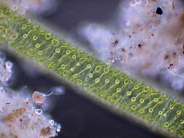
|
|
|
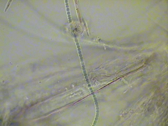
| Anabaena - 40x obj. - COL-D3 |
Anabaena -100 x obj. - COL-D3 |
 |
 |
| Micro-Oligochaete - 100x obj. -COL-D1 + RhFQ |
Larvae of Ephemeroptera - 4x -COL-D3 |
All the COL stops, appropriate to the subject, correctly centred and correctly illuminated, give very marked effects of surface relief, the feeling of an extraordinary depth of field, and very rich shades of colours which differentiate the structure of the subject with a delicacy that the normal bright field illumination does not have.
Paul James insists very much on the centring precautions and the adaptation of the filter and the illumination system to the subject. For the critical cases he has all the reason and you must go through all the needed steps if you want exceptional results.
But in my experience, for most of the usual subjects, one can use the circular oblique illumination with the same freedom that the other diaphragms and stops here discussed and with very good results, leaving the more strict precautions and care for the more difficult materials.
 |
|
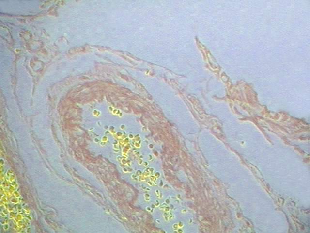 |
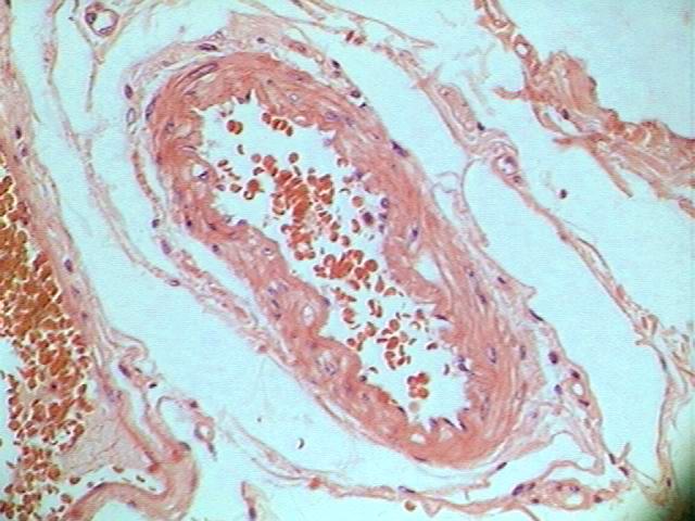
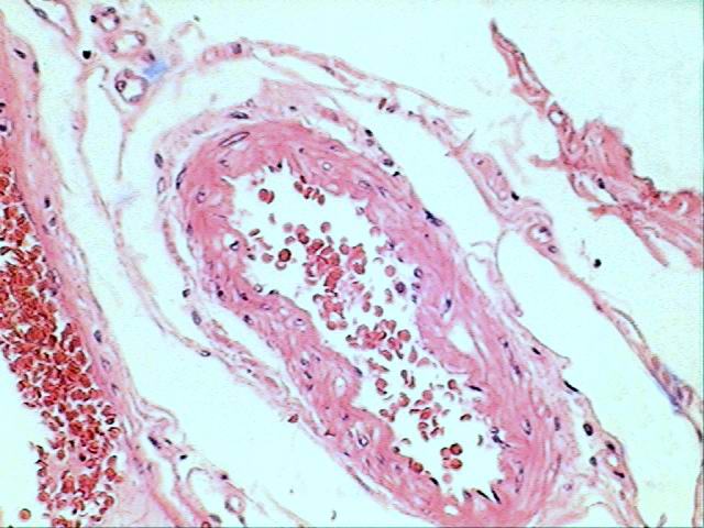
Although
it appears to me only an anecdotal and marginal application, the relief
can be distinguished even in histological preparations coloured with HE
and mounted in Balsam, with a thickness of barely 5 microns, and
various tissues (especially the connective ones) sometimes gives
different brightnesses and colours. Critical illumination - COL-D1 - COL-D3 |
Paul James says correctly, that once the effect of the COL filters has been tried it is very difficult to return to simple bright field illumination. I will add that you will become addict, not only to COL, but to all the filters, diaphragms and stops described here.
THREE IMPORTANT NOTES:
1) The optical effects produced by the COL stops, are not always correctly recorded by all digital cameras (mine is one of those). What one sees under the microscope is much better than the photographic image. Paul James with a camera and equipment many times better than mine, experiences the same problem.
2) It does not matter how beautiful the COL image is. The bright field image always remains the standard. Measurements especially must be made with the images in bright field. The COL gives stereoscopic images, but also creates distortions. You can not get rid of bright field.
3) Nevertheless a very great quantity of details can be only studied, or seen with a greater facility, on the images of the COL diaphragms. It is the same with the images of Phase Contrast or the DIC images.
A LITTLE GALLERY
Here
I add some pictures taken through different devices of the .......
described but of which I have not registered (unforgivable sin) the
used tool.
|
|
|
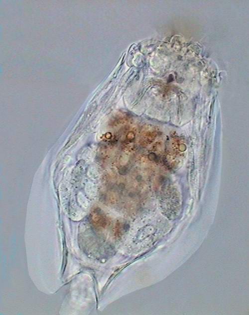
| Euchlanis - a rotifer |
Prorodon - a ciliate |
 |
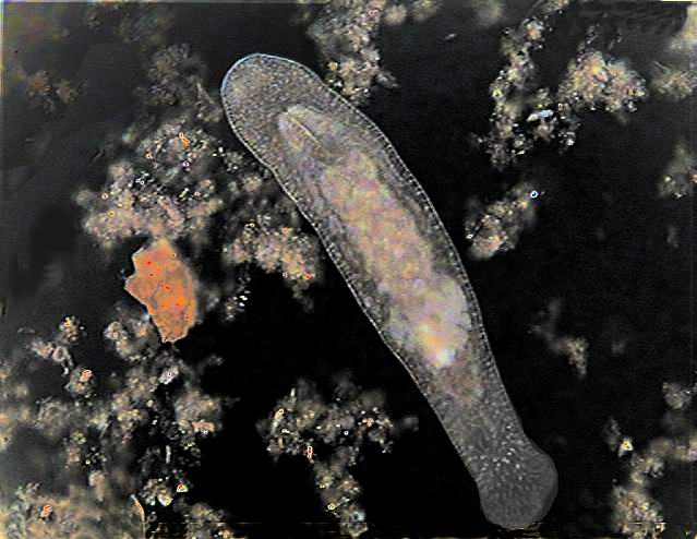 |
| An algae - Spyrogira |
A turbellarian - Macrostomum |
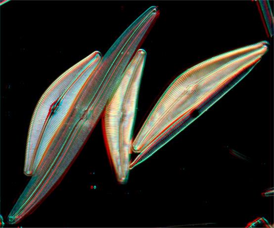
POLARIZATION.- Crystals and many biological materials which have inclusions of various natures gives bright and colourful images if they are examined between crossed Polaroid sheets. There are many publications which detail the applicable methods and show the very beautiful possible results. The technique is very easy; what is difficult is to prepare the crystals. Follow the experts. You can consult
Jean Legrand
Carlos Ratto
www.microscopies.com/DOSSIERS/Magazine/Articles/CR-Cristallisations/Cristallisations.htm
Dave Walker
www.microscopy-uk.org.uk/mag/articles/poldw.html
www.microscopy-uk.org.uk/mag/articles/poldw2.html
FLUORESCENCE.- This advanced technique normally requires a specially equipped microscope, and a very careful technique because the UV rays which promote the fluorescence are very dangerous to the eyes. However there is one work were an amateur summarizes for other amateurs a method which can allow some interesting experiments, if you collect all the materials (filters and fluorescent agents). See
Postal Microscopical Society www.postal-microscopical-society.org.uk
Letter of Keith Brocklehurst, October 10, 1999
EPISCOPY.- Yvan
Lindekens in the message list of the Yahoo "MICROSCOPE" group,
and independently Dominique Voisin in the French MICROSCOPIES Magazine,
had both proposed cheap and easy to do episcopy devices. An
interesting review of the history of the method was presented by
William Ells in MICSCAPE Magazine
Here is the result of my first trial of my first homemade episcope.
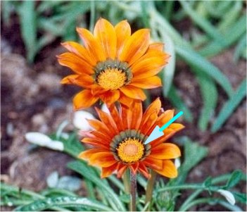 Compound
flower of Gazania sp. ( x 0.5 of natural size aprox.) At right one of
the true flowers from the circle pointed by the arrow . Original is 700
x 827 pixels here reduced to 350 x 413.
|
 |
And why not ANAGLYPHS?
Wim van Egmond described his methods to present photographs in the third dimension
www.microscopy-uk.org.uk/mag/art97b/wim2.html
on the screen of your computer. The easier method is to place on the source of light or in the filter tray a double filter, with a left half red and a right half blue (or green, for another technique). He has presented very beautiful examples on the pages of his web site.
The same technique is used by Giorgio Carboni. He has also displayed some examples.
http://www.funsci.com/fun3_en/hmster/hmster_en.htm
And Daniel Nardin made a very complete presentation in
www.microscopies.com/DOSSIERS/Magazine/Articles/DNardin-Stereomicroscopie-1/Stereomicroscopie-1.htm
The filters are easy to buy or even to make them oneself. One can do some effective ones with two pieces of cellophane. Try the experiment.
You need blue and red glasses to see the anaglyphs. I made mine with cellophane glued onto a cardboard frame which I place over my ordinary glasses. It will be easier for those of you which do not need glasses.
The results are incredible. And after enjoying the anaglyphs of Wim or Giorgio, or yours, you could try a marvellous journey across the Alps (although they are not macro nor micro subjects) at this address
http://perso.wanadoo.fr/alpes-stereo/autresStereo/TDMautresStereo.htm
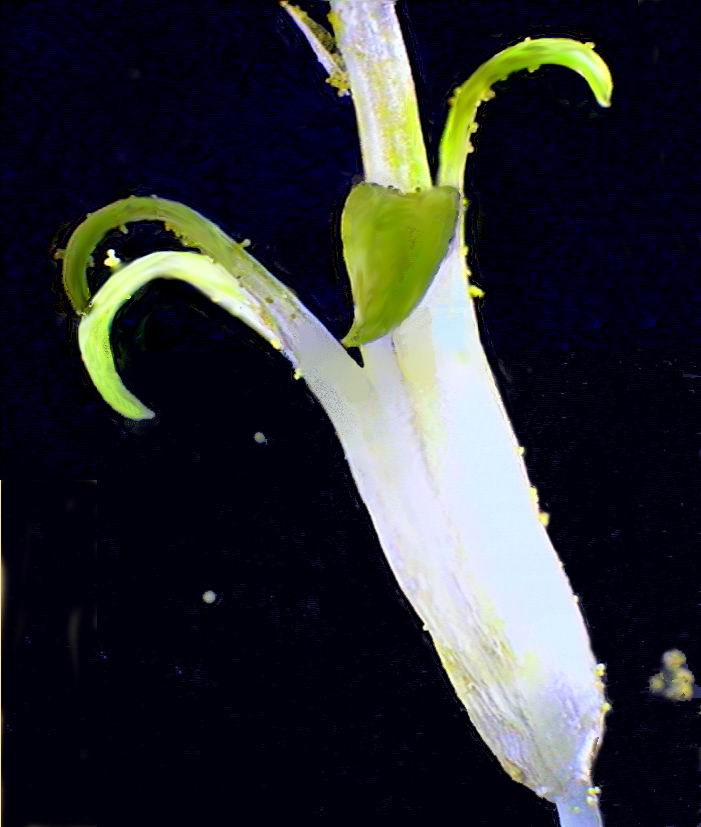
There is another technique to make 3D photomicrographs:
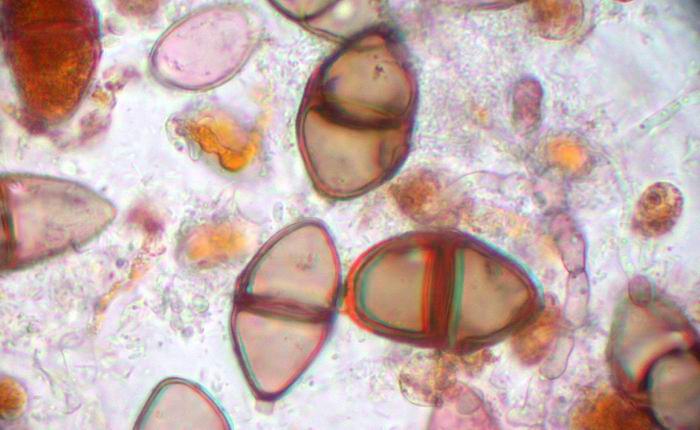
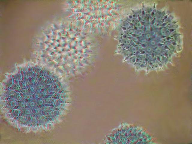
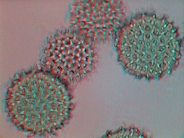
See these tests which I made by using some other's pictures and some personal ones.
 |
The first 3 pictures were treated by a method to produce 3D images from
flat ones. The last one was taken using the Red-Blue filter of Wim van
Egmond and shows the real position of the pollen in the slide. The
original of third picture taken with the COL-D3 stop. |
|
|
|
If you do not have lots of money to buy expensive microscopes with the more sophisticated features, use some time in the manufacture of STOPS, FILTERS AND DIAPHRAGMS. They are not expensive and you can enjoy many happy ventures, while waiting for the cents to accumulate in the small box until the moment comes to acquire 'That One', the long dreamed of MICROSCOPE.
Please report any Web problems or offer general comments to the Micscape Editor.
Micscape is the on-line monthly magazine of the Microscopy
UK web
site at Microscopy-UK