Spicules.
by Mike Samworth
Spicules were common subjects for microslides. Looking
through a copy of the 'Catalogue of Microscopic Objects' from W.
Watson and Sons Ltd., 36th edition, I found seven different
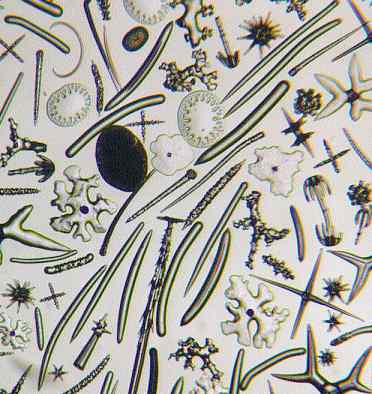
strewn spicule slides offered. The following photomicrograph is
such a strewn slide, showing the numerous variety of spicules
that occur. It is a favourite of mine and I would dearly like to
have such a slide in my own collection.
Group of spicules of sponge, from St
Peter, Hungary (Watson slide).

Of course these slides are very much microscopical objects as
they are only useful to a limited extent in illustrating the
sponge itself. This is because they are isolated siliceous parts
of the horny skeleton of certain organisms e.g. sponges and sea cucumbers. They vary in form greatly,
but whatever the shape or form they serve the same purpose of
strengthening the framework of the animal. When alive, sponges
are composed of a firm, fleshy substance composed of tiny cells.
The horny skeleton is developed in the inter-cellular substance,
and within cells of horny matter these spicula are secreted.
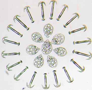
Just as microscopical slidemakers showed their skill and
patience with arranging diatoms, likewise spicules were a
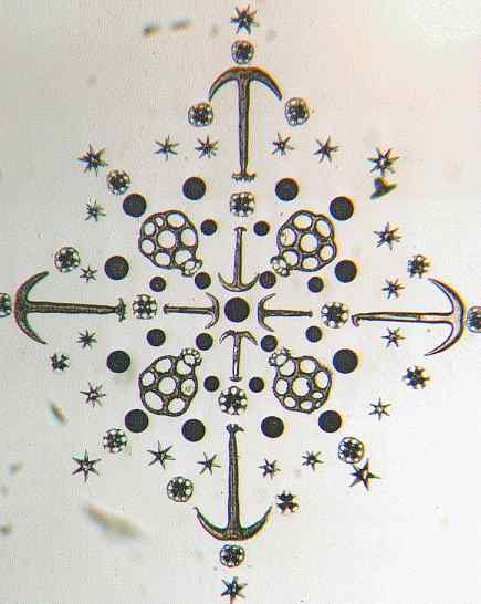
favourite object for arranging into patterns. This is shown in
the following two photomicrographs.
Spicules of Synapta (sea cucumber), Sicily, plates and anchors arranged

I think this feat is exceeded in the next example.
Group of symmetrical Synapta plates (sea cucumber); W
M Gatrell

Perhaps readers who have experience of making such slides
could get in touch to share their knowledge with others, perhaps
even write an article for Micscape.
All photomicrographs by Mike Samworth.
© Microscopy UK or their
contributors.
Published in August 1998
Micscape Magazine.
Please report any Web problems
or offer general comments to the Micscape Editor,
via the contact on current Micscape Index.
Micscape is the on-line monthly
magazine of the Microscopy UK web
site at Microscopy-UK
© Onview.net Ltd, Microscopy-UK, and all contributors 1995 onwards. All rights
reserved. Main site is at www.microscopy-uk.org.uk with full mirror at www.microscopy-uk.net.


