 |
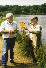
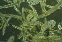
Eric Hollowday examines his catch, and gives some advice to a new member of the club, on how to collect specimens using a net. |
The
Quekett Microscopical Club's trip to Epping Forest.
June 6 1998.
by Steve Durr, UK
The Quekett first made a trip to Epping Forest some time back in the 1870's when Queen Victoria was the reigning monarch of Britain and the empire. Over a century later the Quekett Club are back and just as determined to make the most of the good weather as our forebears did all those years ago. Nine members turned up for what turned out to be a very enjoyable day.
Jeremy Dagley gave us the run of one of the small labs at the Warren headquarters that are used by the staff during working hours. Some members brought their own microscopes and equipment because of the specialised work which they carry out. We set off to visit various ponds which are within easy reach of the Warren centre. We usually spend about 3 hours collecting specimens before retiring to the Robin Hood public house for some much needed refreshment. Eric Hollowday usually keeps every one entertained with his tales of past excursions by Quekett members. Eric joined the Quekett back in the 1940s when the club was very different from what it is today.
|
Eric Hollowday examines his catch, and gives some advice to a new member of the club, on how to collect specimens using a net. |
Ken Jones brought along his video camera and monitor so everyone could see what was happening on the screen, which is very useful if you do not have access to a microscope. A small pond has been built just past the main car park and was full of algae and protozoans. Our attention was immediately drawn to a large group of nodules that were seen attached to some small twigs that had been left in the pond. Upon closer examination I recognised that here was a large colony of Nostoc, which belongs to the Cyanobacteria or Blue Green algae as it used to be known.
In order to get a good photograph of this specimen, it is best to take a very small sample and squash the cover slip very carefully onto the preparation making a thin smear so that it is easier to photograph. Below (left) is a low power view of the gelatinous mass that we found attached to the dead twigs in the pond. Nostoc reproduces by a method called fragmentation, but a special cell called an akinete can withstand desiccation for very many years and still germinate when water is applied. Many species are found on wet pathways or tucked away in the garden, usually somewhere moist like the under edge of the lawn.
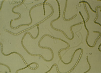 |
The image on the right is a higher powered photograph of the same gelatinous mass taken with a Leica X50 water immersion objective. | 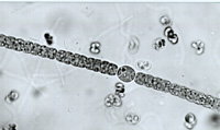 |
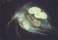
Always a great favourite, is the ever ubiquitous water flea, seen here (below) photographed using darkground microscopy. The brood pouch at the back contains two eggs, and the gut can be seen winding its way through the body, which can be seen through the transparent carapace. Water fleas can quickly decimate any algae present in the water so it is best to filter them off with a small tea strainer.
 |
There is nothing worse that spilling a jar full of pond water that has been left standing for a few days. But for the microscopist, what might be seen as a disaster to someone else, can be an opportunity to get a good photograph, so instead of throwing the smelly water out I decided to put a drop under the microscope and see what might be lurking in there.
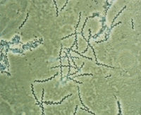
Here are some spirochetes (below) that I photographed using phase contrast. The magnification is about X400 and the corkscrew effect is plain to see in these photographs. Some spirochetes are pathogenic to Mankind and can be found in freshwater. Rats and mice spread one type of spirochete that is called Leptospira, which they pass into the water via their urine. The spirochete can gain access to the body by entering through cuts and grazes that may be found on the body.
 |
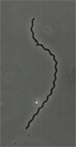 |
| This micro-organism was seen to wind itself into coils and loops. If anyone has any idea what this is please mail me. Again, this is with phase contrast and about x400. | 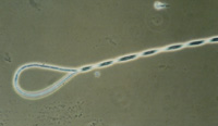 |
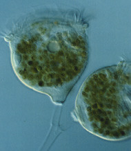
This protozoan (below) was identified for me by Eric Hollowday as Campenella which is a sessile Peritrich and was taken using Nomarski interference which gives a 3D effect to the photograph. These animals hang on to the substratum by a stalk and use their cilia to draw particles of food (mainly bacteria) towards them.
 |
A swirling mass of protozoa (shown below) pulled out of one of the ponds at Wake valley. This photograph was taken with phase contrast and is about X160. They appear to be mostly Spirostomum, which feed on algae and bacteria, they belong to the Heterotrichs.
All in all everyone had an excellent day and it was good to see people enjoying their hobby. One thing about collecting freshwater life, you do have to venture outside and get some fresh air which cannot be a bad thing. Then there is the added pleasure of taking some of the catch back home and trying to identify some of the micro-organisms which you have hopefully caught. Microscopy can be rewarding in many ways, especially on the educational side. Biology, mathematics, physics, optics are all encompassed in the subject of microscopy, and of course computing with its image enhancing capabilities and new publishing techniques have opened up totally new dimensions.
Comments to the author Steve Durr are welcomed.
Editor's note: Visit the Quekett Microscopical Club's website for further information on the Club and benefits of membership.
Published in August 1998 Micscape Magazine.
Please report any
Web problems or offer general comments to the Micscape Editor,
via the contact on current Micscape Index.
Micscape is the
on-line monthly magazine of the Microscopy UK web
site at Microscopy-UK
Please report any Web problems to the Micscape Editor.
Micscape is the on-line monthly magazine of the Microscopy UK Web site at http://www.microscopy-uk.net
WIDTH=1