|
Exploring the Diatom Lab 'Microscope Test Slide in Commemoration of Edmund J. Spitta' with Near UV by David Walker |
Resolving the fine structure of prepared diatom frustules remains a popular aspect of optical microscopy ever since they were found to be of value for the development of objective designs in the 19th century. One of the classic textbooks describing the capabilities of objectives in these studies was Edmund J. Spitta's 'Microscopy. The Construction,Theory and Use of the Microscope' first published in 1907. His photomicrographs are superb and microscopy enthusiasts including myself still aspire to match the quality that he achieved (the 1st, 2nd and 3rd editions published in 1907, 1909 and 1920 are available on archive.org).
Stefano Barone of Diatom Lab has recently offered a 'Microscope Test Slide in Commemoration of Edmund J. Spitta' exhibiting five of the species which Spitta selected. Stefano has shared a splendid article in the December 2022 issue of Micscape presenting images taken with state of the art optics on a Zeiss Axio Imager.A2 compared with results from the typical optics supplied with a 1905 Zeiss Jena 'Jug Handle' Stand IIID—one of the finest stands/optics available in Spitta's time. A state of the art at the time Koritska objective was also used.
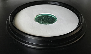
I have been enjoying studying this slide with my own Zeiss Photomicroscope III with 160 mm tube length optics of the period and also on a Nikon Eclipse E800 with modern infinity corrected CFI60 objectives. Both of these microscopes use 100W quartz halogen bulbs that have not been treated to block near UV. This offers a ready means of eking out higher resolutions from standard well corrected brightfield objectives by placing a near UV 400 nm interference filter on the field lens aperture. With an objective still well corrected at 400 nm and with resolution being directly proportional to wavelength, the increase in NA on paper could approach 550 / 400 nm = 1.38X. My past studies with near UV has shown this scaling factor to be roughly borne out with Zeiss 160 mm achromatics and Neofluars.
A hot tungsten wire is a black body radiator and there is a small but finite emission into near UV, increasing as the bulb voltage / filament temp. is increased. The typical monochrome dedicated cameras as used for either astronomy or microscopy are sensitive enough to make use of this emission. A camera is required for all studies but the low emission is perfectly safe - our eyes are exposed to these sort of levels outdoors.
For brightfield LED illuminated microscopes the LED could be changed out for a near UV emitting model. These are typically very powerful and require careful protocols by a continually diligent user to avoid inadvertent eye exposure either by themselves or others. (I do use such LEDs but for transmitted and incident autofluorescence studies.)
One of the benefits of the 100W lamp with filter approach is that all critical setting up of the light train (e.g. Köhler, oblique, darkfield, COL with or without cross polars) can be accomplished in brightfield. The filter can then be put in place while retaining these settings. Replacing a brightfield LED for a near UV model may not necessarily retain these often critical settings of the light train. The filter only needs to be larger than the field iris setting of the lowest power objective planned, 20-25 mm diameter is a convenient size. To maintain camera sensitivity it's best not to pick one with too narrow a value of half bandwith, 10 - 20 nm is suitable. For less sensitive cameras, choosing a filter just into the visible e.g. 410 nm can be used where the emission of a black body rises sharply.
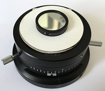
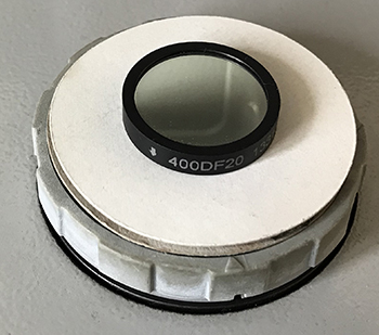
The 400DF20 (400 nm 20 mm diameter) that I use bought from Knight Optical (UK) no longer seems to be available and prices have increased. Some specialist eBay sellers can be checked out, at time of writing an unmounted 25 mm diameter 400 nm with 10 nm half bandwidth is sold by a Chinese dealer for ca. £31 with free shipping which seems excellent value. The closest current Knight Optical equivalent 400DIB25 is £110 plus VAT. A quick look at the Edmund Optics UK range suggest typically twice the price of Knight Optical.


Above. Nikon E800 Eclipse left and middle. The field lens is ca. 45 mm in diameter (left) with field iris some way back towards the lamphouse and controlled by a side knob. A 17 mm near IR blocking filter* does not encroach into the field of the 10X objective, the lowest likely to be used for diatom studies. In practice the de Senarmont DIC base is used which has a 25 mm port (middle).
Zeiss Photomicroscope III (right). An older design where the field lens is 25 mm diameter, with field iris just below the top plate controlled by the outer ring.
For both microscopes with very different optical train designs, a 20 - 25 mm interference filter is suitable.
* A near IR blocking filter is required for brightfield studies with a monochrome camera but not for the interference filter, but in this case a handy support.
The sharp eyed will notice the unidrectional filter arrow for these images is upside down although have not noticed any difference in performance either way!
The images below are a selection taken using either brightfield or near UV with comments in the captions when near UV is of value and when less so. The tonal balance of as-captured diatom images are rather flat so levels adjusted, selected crops resized in Photoshop Elements using Bicubic Sharper. The 100X brightfield images used a green interference filter with a 2X optical relay.
My Nikon E800 is equipped with DIC but it is the dry system, ie the prisms are for NA <1.0. The prisms and condenser top for immersion DIC are uncommon, very expensive and have not invested in them. So the achromatic-aplanatic condenser NA 1.4 oiled was used. Oblique used a card partially covering the interference filter.
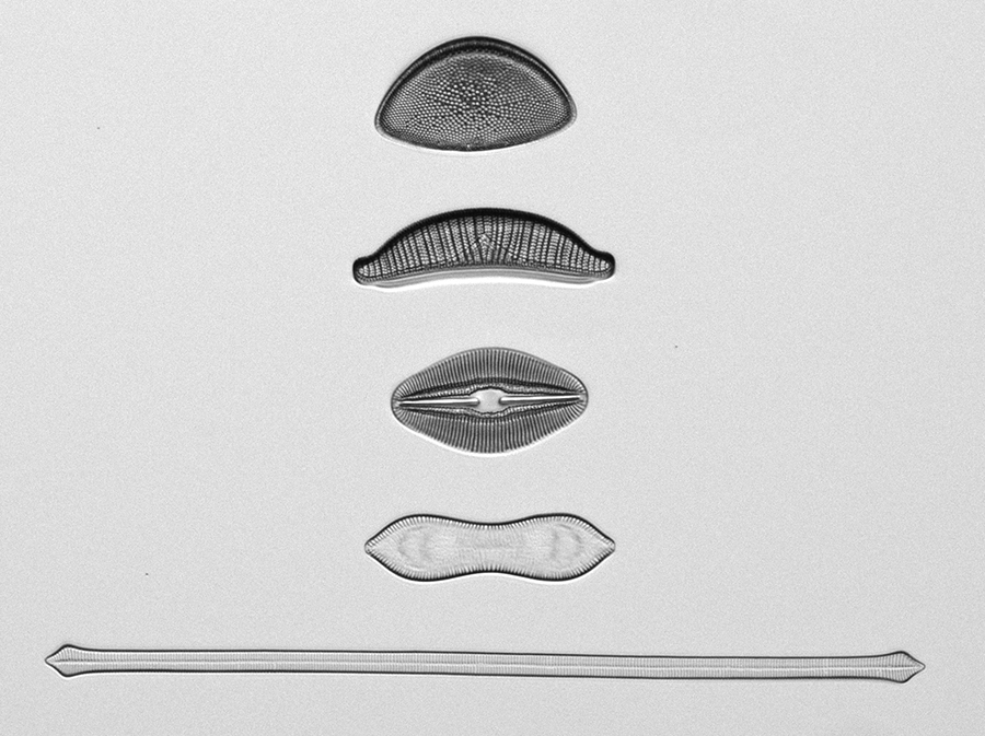
Above. Nikon 10/0.45 planapo brightfield, green filter with some oblique, monochrome ZWO ASI178MM, 1/1.8 inch CMOS 6.4 Mp camera. Near UV is of less value at the lowest mags as the diatoms occupy only a small part of the field where fine detail capture by a modest camera will not readily be seen unless an additional optical mag is added. It can be instructive pushing a low mag/NA objective with camera to the limit by resolving a species like Stauroneis phoenicenteron as described in this Micscape Septemember 2022 article, it was only when upgraded the camera from a 1.3 to 6.4 Mp camera that it was possible with a 2X optical relay and the 10/0.45 objective.
From the top, Hemidiscus cuneiformis, Epithemia turgida, Navicula smithii, Cymatopleura solea, Synedra capitata (slide image rotated 90° to left from as presented on the slide).
Hemidiscus cuneiformis. Spitta remarks that this species requires an NA 0.3 objective to resolve with green oblique light being beneficial. His plate shows resolved with an NA 0.3, 1 inch (ca. 10X). A modern objective of NA 0.45 has no difficulty as shown above. He also notes a 'feint suspicion' of resolution with NA 0.24. I haven't tried it but modern achromatics are typically NA 0.2 to 0.25 so with near UV increasing the NA to ca. 0.28 - 0.34 it would be interesting to compare brightfield and near UV with these objectives.
Epithemia turgida. Spitta discusses the variable appearance of the 'dots', how their appearance varies with the focus plane and their true nature—and notes 'we lean towards the opinion that they should be always shown as circular'. A modern 100X objective suggests they are more rectangular which Stefano Barone showed in his fine image and supported here with a Nikon 100X / 1.3 Plan Fluar in brightfield with polar. Dropping to 400 nm does not seem to reveal any extra fine detail although perhaps some evidence (or artefacts) within the pores. It would be interesting to see an SEM of their true structure. Round (1) has a selection of detailed studies under the SEM but am not entirely clear how some of the figures relate to parts of the diatom. Interesting that the authors note "... areolae often very complicated in structure, making their identity and boundaries difficult to determine ...".
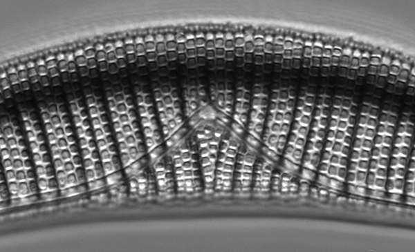
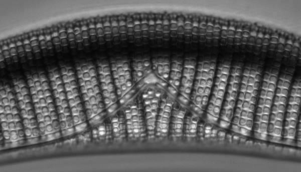
Above, Epithemia turgida central detail, Nikon 100/1.3 Plan Fluar. Upper brightfield, lower 400 nm both slight oblique perpendicular to long axis.
Contrast tends to lower with 400 nm for the same tonal balance adjustments.
Navicula smithii Spitta discusses this species as both a subject for both a low power NA 0.3 and highest power immersed objective. He notes the two rows of dots should be clearly seen. As shown below dropping the wavelength to 400 nm crisps up this detail at the now higher NA.
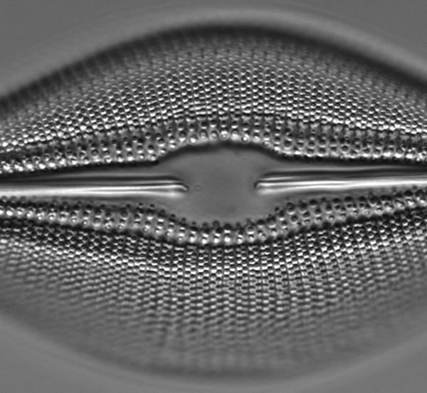

Above, Navicula smithii, central detail, Nikon 100/1.3 Plan Fluar. Upper brightfield, lower 400 nm both slight oblique perpendicular to long axis.
Cymatopleura solea Spitta discusses this species as a subject for the highest powers and NAs (in his optical armoury a 1.5 mm NA 1.35 Koritska apochromatic which Stefano Barone used with the old Zeiss in his article). The fine striae running along the frustule at right angles to the long axis he notes 'are extremely difficult to photograph on account of their great transparency'. By optical microscopy there may be a hint of finer detail in the striae but the SEM in Round (p. 649 plate k) barely shows the finer detail so may be an artefact in the optical microscope. The striae are well resolved in brightfield with slightly crisper detail at 400 nm but the focus planes differ so comparisons are not really significant.
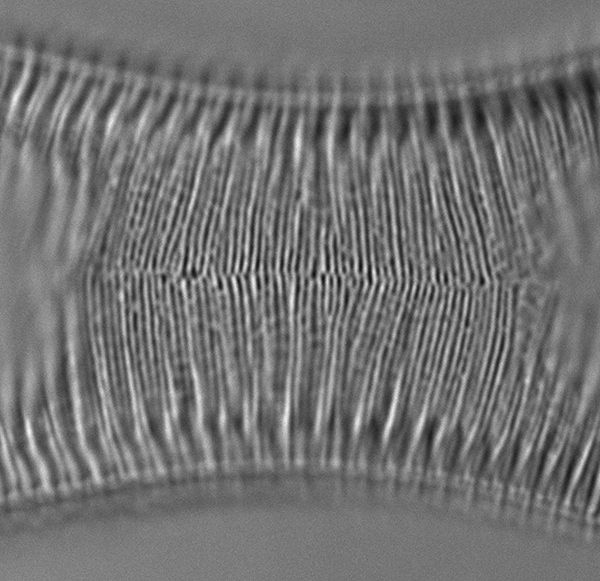
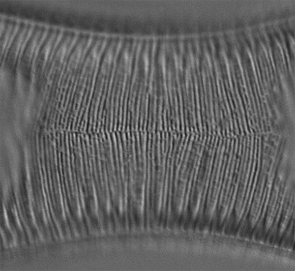
Above, Cymatopleura solea, central detail, Nikon 100/1.3 Plan Fluar. Upper brightfield, lower 400 nm both slight oblique perpendicular to long axis.
Synedra capita The transverse striae are readily resolved at 100X but the aim is to resolve the straie into finer detail. Spitta notes that green light if 'strongly oblique' will resolve but benefits from blue-violet 470 nm. In this example 400 nm crisps up the fine detail somewhat. Orientation of oblique (and DIC) can be critically important to resolve many diatoms and this species is a good example; only the striae are resolved if the oblique is on the long axis of the diatom as shown below. As with many diatom species there are white and black focus planes. Round has SEM images of the striae fine structure for the similar Synedra ulna (p. 370).
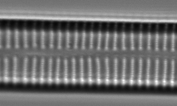
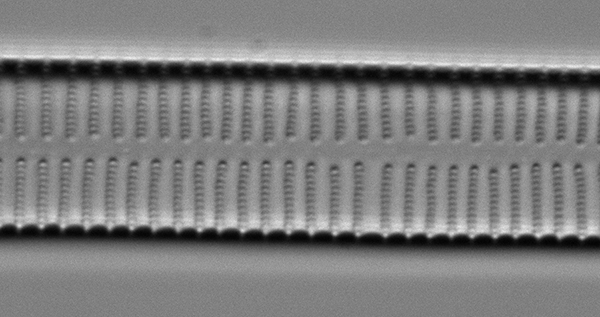
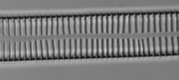
Synedra capita, Nikon 100/1.3 Plan Fluar,white focus plane. Upper brightfield, middle 400 nm both slight oblique perpendicular to long axis.
Bottom, slight oblique parallel to long axis.
General observations The fine structure of species are with appropriate lighting resolved well in brightfield so the benefits of dropping to 400 nm are quite subtle, generally crisping up already resolved detail. This wavelength has a more striking effect for objectives and frustule detail where resolution is either barely achieved or not at all in brightfield. Examples of these will be shared in later articles using the Diatom Lab Test Slide 2.0 and a selection of Diatom Lab's selected single species diatom test slides.
Reference
1) F. E. Round, R. M. Crawford, D. G. Mann, The Diatoms, Biology & Morphology of the Genera, Cambridge University Press, 1990. (I'm fortunate to have the hardback, the images in the softback are noticeably inferior.)
Published in the August 2023 edition of Micscape.
Please report any Web problems or offer general comments to the Micscape Editor .
Micscape is the on-line monthly magazine of the Microscopy UK web site at Microscopy-UK
©
Onview.net Ltd, Microscopy-UK, and all contributors 1995
onwards. All rights reserved.
Main site is at www.microscopy-uk.org.uk.