|
by Roland Mortimer, Brazil |
Having never seen a mosquito in England, I'd always thought of the 'bite' being performed by a needle-like piece of apparatus. This all changed when I came to Brazil, waking up in the mornings and finding lumps on my skin, could these be flea-bites I thought in my ignorance.
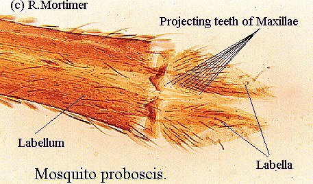
They were actually mosquito-bites, what I didn't know was that I was very allergic to the saliva which caused the lumps, some of them very swollen and painful. Now, after having been bitten more times than I'd like to remember, I seem to have anti-bodies and the swellings don't occur anymore. I decided to take a look at this terrible piece of weaponry under the microscope, a lot of patience and a steady hand are required to remove the biting parts from the labium or outer sheath, the whole thing is finer than a human hair and the internal apparatus even finer still.
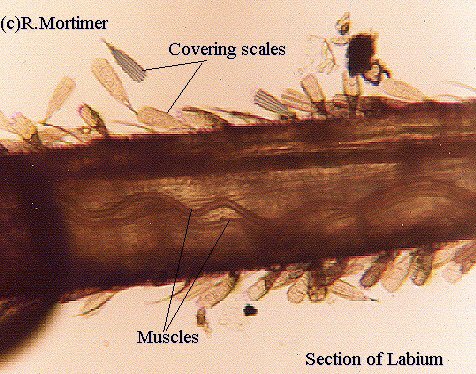
Once removed and observed at a magnification of x200 one can see a long narrow piece of material with parallel sides tapering to a point at the tip. This 'flat' strip is in fact hollow and is made up of several parts, the labrum-epipharynx, the mandibles (2), hypopharynx, through which the salivary duct runs, and maxillae (2) i.e. six parts in all. The muscles and trachea are contained within the labium, or sheath. On biting, the labella are pressed against the skin and thus act as a guide to the piercing organ, the labium buckles and it too acts as a guide, none of these parts enters the resulting wound.
 The
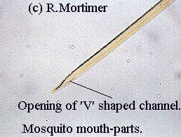
labrum-epipharynx is composed of two lamellae which form a 'V'-shaped channel
with ventral opening along its length ending in a sharp point. Fitting
closely to the ventral surface of the labrum-epipharynx is the hypopharynx,
a thin chitinous lamella, if you can imagine the 'V' shaped channel closed
on the opening of the V by this thin flat plate then you will clearly see
this forms a tube through which the blood is sucked.
The
labrum-epipharynx is composed of two lamellae which form a 'V'-shaped channel
with ventral opening along its length ending in a sharp point. Fitting
closely to the ventral surface of the labrum-epipharynx is the hypopharynx,
a thin chitinous lamella, if you can imagine the 'V' shaped channel closed
on the opening of the V by this thin flat plate then you will clearly see
this forms a tube through which the blood is sucked.
In the thicker chitinous portion of the hypopharynx runs a fine channel from base to tip, this is the salivary duct through which saliva from the mosquito's salivary glands is forced into the wound, the saliva contains anti-coagulants, probably the initial cause of my 'lumps'. The mandibles are very delicate structures which terminate in a sharp point, these lie at the side of the hypopharynx.
 The
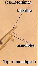
maxillae are much more robust in structure and terminate in sharp points
too, the tapered points being lined with a row of backward pointing teeth
much like a saw. The teeth cause more damage once inside the delicate tissues
below the skin and thus cause a greater haemorrhage than would a straight,
smooth, needle-like appendage. These piercing mouth-parts belong only to
the female, for it is only she who needs blood protein to mature her fertilised
eggs , the poor old male has to make do with fruit juices or flower nectar
because his mouth-parts are not as well developed as the females', or the
mandibles and maxillae may even be totally lacking.
The
maxillae are much more robust in structure and terminate in sharp points
too, the tapered points being lined with a row of backward pointing teeth
much like a saw. The teeth cause more damage once inside the delicate tissues
below the skin and thus cause a greater haemorrhage than would a straight,
smooth, needle-like appendage. These piercing mouth-parts belong only to
the female, for it is only she who needs blood protein to mature her fertilised
eggs , the poor old male has to make do with fruit juices or flower nectar
because his mouth-parts are not as well developed as the females', or the
mandibles and maxillae may even be totally lacking.
Comments to the author Roland Mortimer welcomed.
Please report any Web problems or offer general comments
to the Micscape
Editor,
via the contact on current Micscape Index.
Micscape is the on-line monthly magazine of the Microscopy
UK web
site at Microscopy-UK