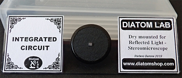
Exploring the Diatom Lab prepared slide of an integrated circuit chip.
by David Walker, UK
Like many amateur microscopists I find the detail of integrated circuits fascinating. They are an excellent subject to try various incident lighting techniques whether on a compound microscope or stereo. My own attempts to release a chip from its package have been mixed. Michael Reese Much describes a protocol for some types of chip but requires chemical procedures which as he describes requires great care. So I treated myself to a prepared slide from Stefano Barone's Diatom Lab sold on eBay under their 'diatomshop' eBay ID (currently £22 Buy it Now). It is impeccably prepared / mounted and wire contacts have been removed to present a flat uncluttered surface. Sensibly it does not use a coverslip so can be kept dust free in the slide box supplied.

Incident light - Zeiss Photomicroscope III epi system.
I treated myself to the epi head unit some years ago and later two DIC objectives, a 4/0.1 and 40/0.85. Epi DIC is more cost effective than transmitted as the prism above the objectives serves two roles both for incoming and outgoing light. I don't have a wealth of suitable subjects to use with them but ICs are ideal. How the DIC bias retardation and angle of subject to the shear axis affects the detail presented can be explored. DIC is the only time I use on-axis epi lighting as find this type of lighting very flat for standard incident light studies and much prefer off-axis lighting with either a gooseneck light or small LEDs on gooseneck powered by USB dock which can be used for quite close working distance objectives.
For fellow folk like me who find any sort of stacking tedious(!), they are also useful subjects to cut our teeth as only two or three image planes need to be focussed to capture the planes of circuitry.
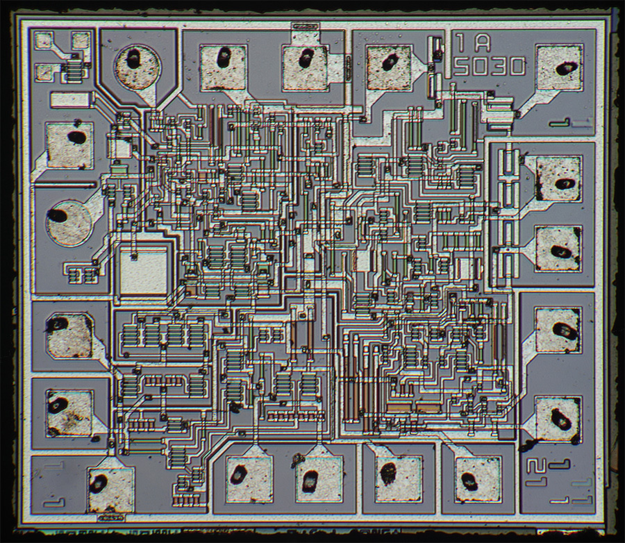
The whole chip, Zeiss 4/0.1 DIC epi objective. This example measures 1.56 x 1.38 mm. Two image stitch using the Panorama feature in Photoshop Elements.
The chip sits neatly in the circular field of my 10X KpL eyepieces with the 4X epi objective but not quite my camera setup using a Canon 600D DSLR with APS sensor.
This sort of subject can be a good test to assess both geometric and optical distortion.
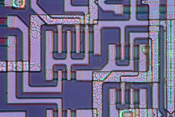
Zeiss 40/0.85 DIC epi objective with lambda waveplate. Stack of three images in CombineZP.
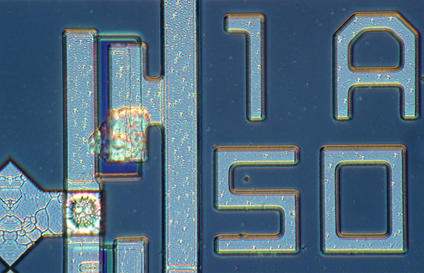
Zeiss 40/0.85 DIC epi objective with lambda wave plate. Stack of two images in CombineZP.
Stereo microscope
As the label suggests, it is also an excellent subject for the stereo where lighting techniques can be explored to see if the subtle surface detail can be enhanced.
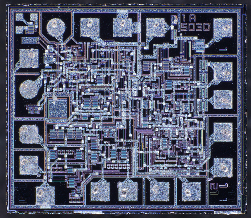
Leica S8 APO at 80X max using a Sony NEX 5N body. 144 LED ringlight. The slide was angled at six degrees to the photo side of the Greenough optics so that the subject was planar with respect to the optical axis.
The linear features provide good practice for taking measurements. The tracks are typically ca. 0.02 mm wide in this relatively simple circuit and much wider than the now nanometre level tracks of the high density chips typical of modern computers.
In my view the slide is excellent value for money and maintains the high standards set by the Diatom Lab shop (I have a number of the diatom slides). It has proved to have a number of valuable roles as a subject on both the compound and stereo microscope. It would also be a good subject to explore with extreme macro techniques where objectives on bellows or other approaches are used. Some specialist extreme macro websites use integrated circuits as test subjects as their regular flat features can be used to spot geometric and optical distortion. In fact, it has given me an idea to try extreme macro myself with this slide to compare the results from using the microscopes.
Comments to the author are welcomed.
Published in the April 2020 edition of Micscape.
Please report any Web problems or offer general comments to the Micscape Editor .
Micscape is the on-line monthly magazine of the Microscopy UK web site at Microscopy-UK
©
Onview.net Ltd, Microscopy-UK, and all contributors 1995
onwards. All rights reserved.
Main site is at
www.microscopy-uk.org.uk.