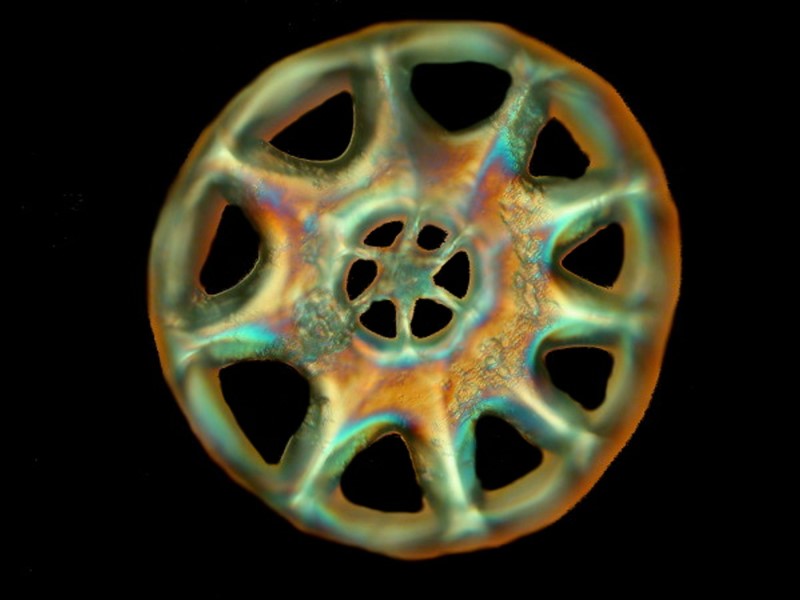
What look like a thin, pinkish-white 6 inch worm, burrows in the sea bottom, ingest sand and other detritus, has a ring of tentacles around the mouth, has white warty skin, and has some especially odd bumps covered by a very thin membrane which encloses a clump of spicules which look like tiny glass wheels with spokes? You guessed it–Chirodota. Chirodota is one of the odder members of the holothuroids or sea cucumbers, although when I stop and think about it, almost all of them are fairly odd.
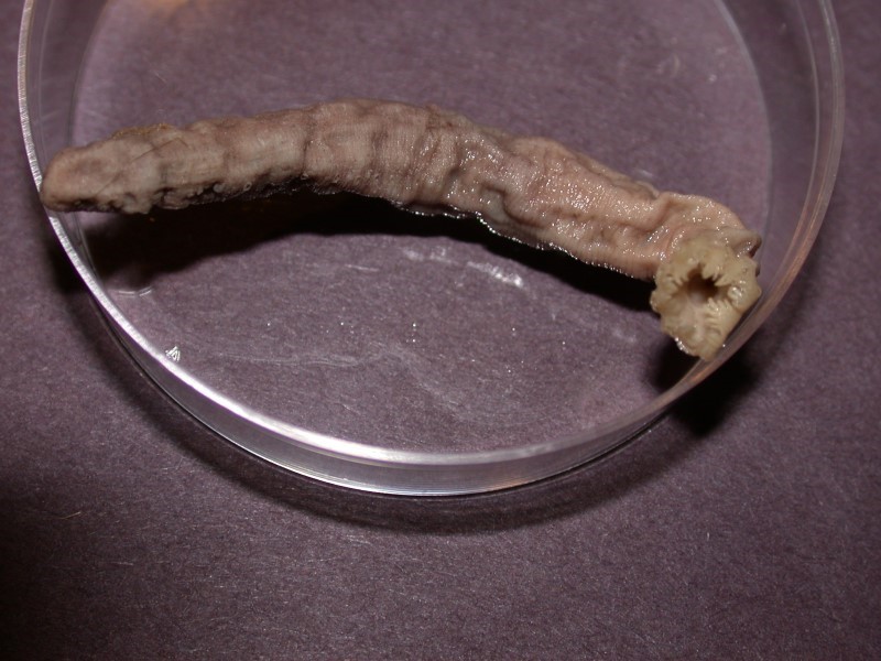
Note: There is some dispute over whether it’s name is “Chirodota” or “Chiridota”; I am using the former.
Chirodota, like many cucumbers will, when seriously disturbed, eviscerate, and even worse, may seriously rupture, essentially disemboweling itself. When placed in a killing and preserving fluid having been previously anesthetized, even then, perverse creatures that they are, they may perform this holothurian hara-kiri anyway, completely oblivious of their obligation not to behave in such a fashion under anesthesia. As a consequence, the preserved specimens which I have are in some disarray but, nonetheless, are extraordinarily interesting. Many people think that these creatures are remarkable ugly, but take a gander at the spicules;--how can you dismiss an organism when it can produce such an elegant structure.
Furthermore, because sea cucumbers function as the “vacuum cleaners” of the sea, their intestines often contain the remains of diatoms, forams, radiolaria, and bits and pieces of larger creatures whose calcareous or chitinous shells have been broken up by larger predators.
My first encounter with Chirodota was a specimen which a marine biologist in California kindly sent me. For many years, I kept it intact since it was the only such specimen I had and it was quite well-preserved. I did, however, early on, notice what appeared to be a few clumps of sand grains toward the anterior end and I got curious and scraped one off. Imagine my surprise when, under the microscope, I found tiny, calcareous wheels with spokes. Still, I wanted to keep the specimen intact for reference. Then a few years ago, while sorting a miscellaneous shipment of specimens, I happened across several more specimens of Chirodota. Recently, I selected 2 fairly mangled and eviscerated specimens for examination. Their condition was likely the result of being plunged directly into formaldehyde.
I decided that before dissecting them, I would first remove the clumps of spicules and keep them separate. In the first specimen, as in the reference specimen, the clumps seemed to be restricted to the anterior end and located under a very thin layer of tissue, so that once I caught on to their appearance, they were fairly easy to locate. So, I began to form an hypothesis–the clumps occur primarily, if not exclusively near the anterior end. Wrong! In the second specimen, I not only found clumps in the general anterior region, but immediately below the ring of tentacles at the “head”, on the “neck”, along the main part of the body, and even at the extremely posterior tip. So much for my hypothesizing. Even more surprising, I also found a number of clumps under a very thin layer of tissue on the outer layer of the interesting beasties, but clearly well within the organism itself! These “clumps” as I have been calling them are very interesting indeed. At first, when I went to remove a clump for microscopic examination, I thought that once I punctured the skin where they were located, I would have tiny wheels rolling all over the place. But, as it turns out, the spicules come in little packets, as if they were all neatly bundled in a tiny envelope of plastic food wrap. I haven’t yet been able to determine whether there is a very thin membrane encasing them or whether there is some sort of mucous or “glue” that keeps them packaged. At the moment, I am leaning toward the latter explanation or it could be both, but don’t rely on me–remember my earlier wrong hypothesis.
Certainly something causes them to adhere in clusters. When I took a pair of micro-dissecting needles and tried to separate the wheels, I experienced significant frustration. They simply wouldn’t separate. So, the next step was chemical warfare–sodium hypochlorite (household bleach). A drop of two on a side immediately attacked the “glue” producing many small bubbles, making the process virtually impossible to observe. After a few minutes to half an hour, the separation is largely accomplished. During this period, it may be necessary to add either more sodium hypochlorite or a drop of water from time to time. If you permit the slide to dry, you will get a clump of crystals. [CAUTION: Sodium hypochlorite is caustic and, as you observed on the slide, will attack tissue. Be especially careful never to let any such caustics get into your eyes, since they can do irreparable damage. Also, never mix these with other chemicals because certain reactions will produce deadly chlorine gas.]
Once the spicules are separated out on the slide, then comes the rather tedious task of multiple rinsings. Using a micro-pipet, remove the bleach solution (or as the French call it: Eau de Javelle–they manage to make even unpleasant things sound nice–which is perhaps why, for so long, French was the language of diplomacy). As you remove it, take care not to remove any of the spicules. Pipet off the water (distilled), discard it; add more, discard it, and after 734 repetitions you can feel fairly confident that all the bleach solution has been removed. Well, perhaps you don’t need quite that many rinses, but 10 or 12 is certainly better than 3 or 5. All of this can be done with less fuss and bother by carrying out these procedures in small tubes or Petri dishes, because you don’t have to worry as much about evaporation and pipetting the solutions is easier. Take care to stopper the tube in such a way that oxygen being released can escape as the bleach attacks the tissue. A small ball of cotton is quite effective. If you use a cork or rubber stopper, the pressure may build up enough to blow them out of the tube spattering caustic solution on you and/or your equipment. Just be sure that you always allow sufficient time for the spicules to settle to the bottom and then, when you do the pipetting, angle the tube a bit and remove the water slowly until you have only about 1/4 of an inch remaining in the bottom. Also, when using tubes, you can now add the distilled water by means of a wash bottle, thus agitating the spicules and helping to remove any deposits on them. Be sure, however, to do this enough times. How can you tell? When you think that the spicules are clean, take a tiny drop from the bottom including just a few spicules, place it on a clean slide, let it dry. Examine the slide carefully under a compound microscope, at at least 100x, and you may wish to go even higher. If you don’t find deposits on the spicules, then you can store them for mounting, either in distilled water (preferred) or a neutral pH high-grade alcohol, but with the latter you need to be aware of the potential for evaporation problems.
It’s worth going through the slide procedure at least once, just to observe the emergence of the marvelous “wheels” from a mass of bubbles but, after that, you’ll very likely prefer the tube or small Petri dish method. However, in all fairness, I should warn you that these particular spicules are quite fragile. These procedures can be applied to a wide variety of calcareous structures in invertebrates.
Chirodota presents special problems and aggravations. As I mentioned, the spicules are fragile and I now strongly suspect that the bleach etches them and thereby weakens them in such a fashion that multiple rinsing leaves few of the “wheels” completely intact. Further more the bleach is not fully effective in removing the bits of organic material surrounding the spicule clusters nor, seemingly, does it remove all of the vestiges of the “glue” that holds the spicules together in packets.
I discovered this latter point when, after multiple rinsing, I tried to transfer the remaining spicules to a Petri dish for observation only to find that they were tenaciously stuck to the plastic tube. Would glass tubes work better? I doubt it.
My next step will be to try a concentrated solution of hydrogen peroxide (35%). The solution you buy at the store is 3%. I will very likely begin with a 10% solution and work up if need be. [CAUTION: Concentrated hydrogen peroxide is a powerful oxidizer and can produce very dangerous results if mixed with the wrong chemicals.] I have also ordered some Pancreatin which is a digestive enzyme. Some work has been done using this substance to remove organic material surround the spicules and this approach seems promising. Both optical and SEM examinations indicate that Pancreatin does not produce etching as does sodium hypochlorite.
In addition to the spicules, Chirodota has, just inside the oral cavity, a series of calcareous plates, which form the circumoral ring. There are 10 such plates and these too can be isolated and mounted, although their form is not as appealing as that of the spicules. This ring helps provide the support for the powerful muscles which extend and retract the tentacles.
Chirodota is an indiscriminate feeder and simply shovels in virtually anything within reach of its tentacles. When I dissected the two specimens I had been removing spicule packets from, I was amazed to find that the stomach and long looping intestines packed full of sand. The density and size of some of the individual sand grains make one wonder why the intestinal walls don’t rupture, for they are quite thin, but apparently both strong and elastic. The sheer amount of the sand seemed hardly credible to me and I venture to say that in both volume and weight, it constituted more than 52% of the body mass. (However, you need to remember that 73.9% of all statistics are pure fabrications.) In my initial examinations, I was able to find in the stomach and intestines, sand, 2 forams, sand, 2 fragments of worm tubes, sand, 1 large centric diatom, sand, sand, and sand. So, I took some of the debris and put it on slides to scan at high magnification. Even at 400x, evidence of anything much in the way of sustenance was meager. There were some very small diatoms, small algal cells and bits of algal filaments, and a few sponge spicules. And, to my considerable surprise, there in all that stomach debris was an anchor! Not a ship’s anchor, but a spicule much prized by Victorian micro-mounters which comes from a genus of sea cucumbers called Synapta. Had I been a 19th Century sailor and known about these spicules, they might indeed have made me very superstitious–spicules in the form of anchors! The appearance of one here does not mean that Chirodota feeds on synaptids, but simply that a synaptid succumbed in the area and some anchor spicules got deposited in the debris this Chirodota was feeding on.
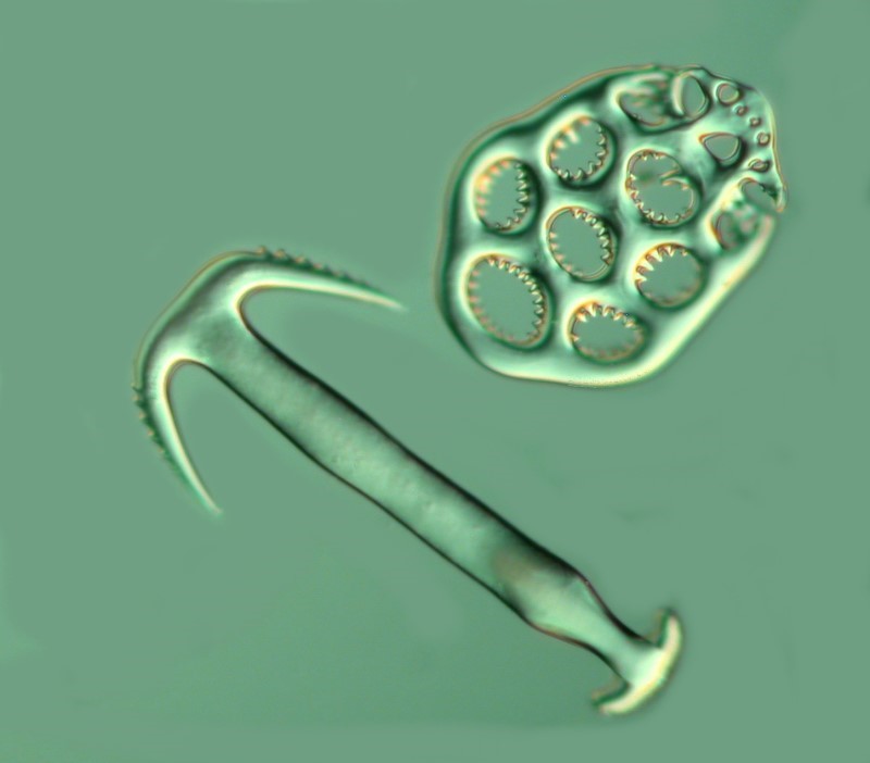
I began to wonder if these bizarre little creatures had found some magical way of converting quartz into food and if I could figure out how, then I could certainly win a Nobel Prize by eliminating starvation. However, as soon as word of my discovery leaked out, some greedy poltroon would buy up all the deserts and start selling sand for $100 a pound.
How can a creature 5 or 6 inches long (exact length is difficult to determine, since these organism are quite elastic) and possessed of a diameter varying from ½ to 1 inch, extract enough nutriment from of this sandy debris to survive and why didn’t all of these sharply pointed sand grains puncture the intestine walls?
As I dissected these 2 specimens, I observed that the area around the tentacles and extending back to the region of the stomach was possessed of remarkably strong and thick musculature. Further on, in the intestine, the walls were significantly thinner, but highly elastic and no ruptures were apparent. So, what’s going on? Clearly with all of this elastic musculature, undulating waves are moving significant amounts of debris through Chirodota’s system in rather short order. Furthermore, the heavy musculature at the anterior end must produce a powerful grinding effect as the sand grains and other debris are moved toward the stomach. Such action would use the sand grains to virtually pulverize everything else which would explain why only a few spicules, very small diatoms and forams, and bits of cellulose algal envelopes are detectable in the digestive system. I would also suspect that the digestive capabilities of Chirodota must be quite remarkable since, for its size, it needs to be extremely efficient at metabolizing any organic material that comes its way.
I am, however, still puzzled by why Chirodota has these odd little self-contained packets of spicules randomly distributed around its body. It certain can’t be as a deterrent to predators, since the gut full of indigestible sand is a much more likely deterrent than a few little packets of calcareous wheels. Perhaps Chirodota as it dozes after a gigantic repast of debris, dreams of spoked wheels and creates little caches of them in its own body. We don’t yet know much about how it creates these wheels and we certainly don’t know why it produces wheels rather than squares or triangles or spheres since such shapes do indeed appear among the diatoms. Other species of sea cucumbers produce a variety of differently shaped spicules including perforated plates, the anchors mentioned above, and boomerangs punctured with holes–but Chirodota, in my experience, produces only wheels.
Eupentacta has an epidermis composed of masses of minute spicules numbering, I would venture, in the tens of thousands in a single specimen–a veritable calcareous armor. Certainly some predators would be deterred by a mouthful of such grit, but I would think that the clever little Chirodota’s distribution of densely packed sand grains through it’s length would be even more of a deterrent. So, the spicules are probably just an evolutionary vestige, but one which, nonetheless, can bring us aesthetic pleasure as Victorian slidemakers recognized.
Some sea cucumbers have no spicules at all. So, is this evolutionary “progress”?–less is better? Many biologists have argued that this is the case. The marvelous heavy pink shells of the giant conch or the enormous Tridacna have often been discussed as being indications that these organisms are, on the evolutionary scale, “lower” than their less encumbered relatives, such as, squid and octopi. Not being a biologist, I sometimes wonder if such judgments aren’t anthropomorphic projections. It is almost inevitable that evaluative and hierarchical aspects tend to sneak into our conceptualizations.
We assert that it’s more efficient to swim and maneuver like a squid than to drag a whacking great shell around like a conch does and so the squid is “more evolved”, “higher” on the taxonomic scale. However, we need to remind ourselves that efficiency is a human criterion. Mollusks have been around for hundreds of millions of years and both the sleek fast cephalopods and the lumbering heavy-shelled gastropods still survive. If we were to use the criteria a versatility and maneuverability in terms of swimming, they perhaps chaetognaths (the “arrow worms”) might win first prize.
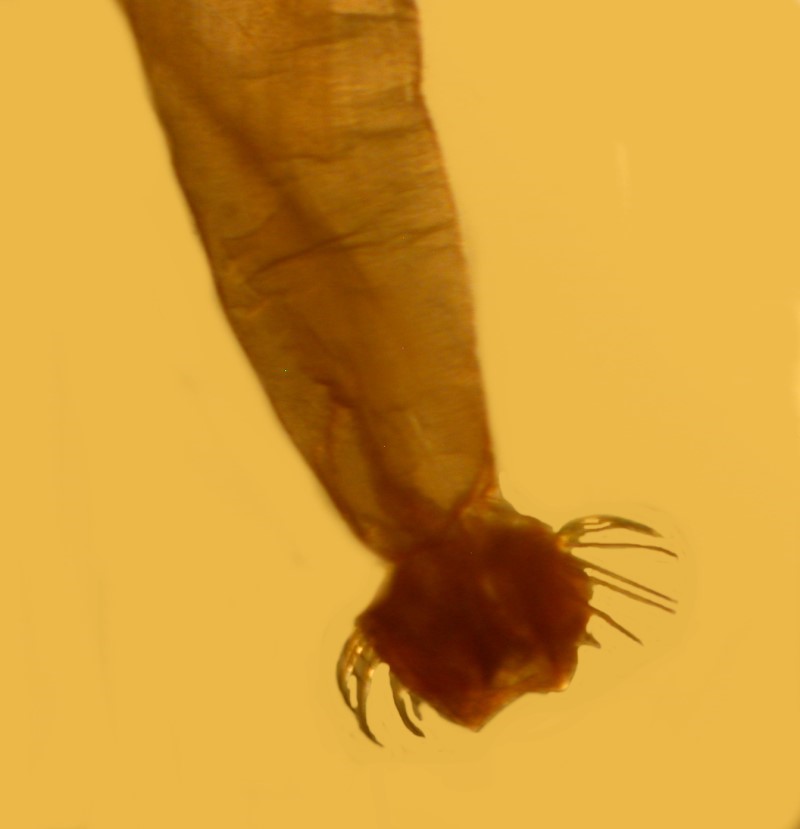
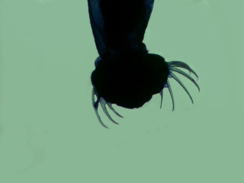
They might almost be described as the sharks of the planktonic world. They are sleek, lightening-quick and beautifully “designed” hydrodynamically. They also have an insatiable appetite (a trait which this portly, retired professor finds charming). But these creatures are “lowly” on the phylogenetic tree in relation to cephalopods or even conchs. Further up, we find the urochordates, such as, ascidians or “sea squirts”, some of which are so undistinguished in appearance as to be readily mistaken for sponges. Yet, in their larval stage, they have a notochord or the beginning of a “backbone”. When they become sessile (attached), they lose this structure. Ascidians are strange and fascinating creatures, but there are other “lower” invertebrates that exhibit more complex morphology overall. So, in our hierarchical schemes, having the beginning of a backbone, even temporarily, places them higher up the evolutionary ladder. In terms of a nervous system, ascidians aren’t going to win any prizes. The brain is a tiny little clump with a few ganglia; cephalopods have much larger and quite sophisticated brains. Perhaps the biological sciences need a theory of taxonomic or phylogenetic relativity. If we use survival as the primary criterion in an evolutionary hierarchy, then surely certain species of bacteria and viruses are terrifyingly efficient–that criterion that we humans prize so highly. The extraordinary ability of these “primitive” organisms to mutate and adapt to adverse conditions has to impress us, even though we recognize the appalling havoc which they can wreak on human communities.
However, being the egotistical organisms that we are, it is the survival of the individual that is of primary concern to us. Giant Sequoias and bristlecone pines have us outclassed by far in terms of longevity, but even in the world invertebrates there creatures, such as, rotifers and tardigrades, which can go into a state of suspended animation and be “revived” over a century later, thus rivaling our own lie spans. A pretty good trick for a “primitive” animal.
How does all of this connect of Chirodota? Well, given its ability to adapt to its rather stark and somewhat specialized environment, it is really quite efficient. Furthermore by burrowing, it increases its security and by having its gut constantly filled with sand, it doesn’t qualify as a tasty morsel for most predators. We need to remind ourselves occasionally that our classificatory systems are conceptual tools which we construct in order to help us understand the bewildering complexity of the countless number of creatures with which we share this planet. We need to be clear about the criteria we use in making judgments about “higher” and “lower” and be cautious not to read large-scale “purposiveness’, design, or progress into evolutionary processes, because that tends to lead to a kind of dogmatic teleology which additionally, leads, all-too-readily away from the realm of science into the misty vapors of theology.
There are those who wish to argue that the marvelous wheel spicules of Chirodota are evidence of the existence of a God and it is further argued that the design of the extraordinary glass tower of the sponge Euplectella (the Venus Flower Basket), the spirals of Nautilus shells and galaxies, the remarkable symbiosis between protists and termites, and the patterns of snowflakes are all evidence of some Grand Designer, who generally just happens to turn out to be male, Caucasian, and Christian. The ancient Greeks asserted that nature causes us to experience thauma–wonder, astonishment, amazement–and certainly Chirodota’s little wheels are wonderful, astonishing and amazing, but hardly grounds for positing the existence of Zeus, Quetzlcoatl, Osiris, or Jehovah. We need to learn to enjoy and respect our ignorance as we study nature to discover further truths, rather than using it as an excuse to create mythical beings with fantastical powers.
A Carrollingian (sic) Addendum
Regarding Eating and Busyness
In many human cultures, constant activity has been regarded as a virtue and celebrated. Popular lore and poets have created the image of constant industry around the bee, but at night bees return to their hives and catch at least 40 winks. On summer mornings at 7:00 when the air here at 7,200 feet is still only 40 or 45 degrees Fahrenheit, I have found bees contentedly napping on hollyhocks and sunflowers, whereas the sea cucumbers are down in their chilly depths still churning away.
Isaac Watts in his poem “Against Idleness and Mischief” from his 1715 book of Divine Songs for Children tells us:
“How doth the little busy bee
Improve each shining hour,
And gather honey all day
From every opening flower!
How skillfully she builds her cell!
How neat she spreads the wax!
And labours hard to store it well
With the sweet food she makes.”
(There are two more stanzas which you can look up for yourself if you want to read the riveting conclusion.)
However, I suppose it won’t do to change it to:
“How does the little busy sea cucumber...”
although Lewis Carroll had some fun by substituting a crocodile:
“How doth the little crocodile
Improve his shining tail,
And pour the water of the Nile,
On every golden scale!
How cheerfully he seems to grin
How neatly he spread his claws,
And welcomes little fishes in,
With gently smiling jaws!”
But back to the busy sea cucumbers. Again we find some puzzles, for some species of Stichopus (a rather large holothurian), after breeding, may go into a dormant non-feeding stage for 2 or 3 months. Some holothuroids are over 6 feet long, move slowly, and take substantial rest breaks, so once again we have a reminder that what humans sometimes see as virtues in ourselves are blatantly ignored by other creatures.