Stars of the Marshes
by Wim van Egmond
The last Friday of May this year the Dutch
Microscopy Club NVVM had an excursion to the fens of Oisterwijk.
Host of this trip was one of our fellow members Frans Roefs who
lives near the location. He is an amateur microscopist like most
of our members, but by chance he became involved in research
carried out by Natuurmonumenten, the owner of these fens.Their
aim is to monitor the different small lakes and find out how to
restore the original ecosystem as well as possible. Because Frans
is studying the lakes and knows the area very well he was the
perfect guide. He could show us the most interesting spots.
 That
day we had a good catch with several kinds of interesting algae.
Most prominent were the desmids. There were all kinds of Closterium,
Cosmarium, Staurastrum and a few Micrasterias
species in our finds. For me this was the right occasion to make
sketches of the several species of Staurastrum.
That
day we had a good catch with several kinds of interesting algae.
Most prominent were the desmids. There were all kinds of Closterium,
Cosmarium, Staurastrum and a few Micrasterias
species in our finds. For me this was the right occasion to make
sketches of the several species of Staurastrum.
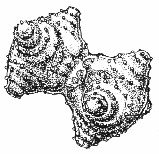
About the images.
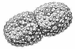
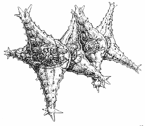 Staurastrum is a particularly interesting
organism to make drawings of because it's so tiny. That makes it
very difficult to photograph. To see the fine details you should
use a 100x immersion lens. The image then presented has such
shallow depth of field that it's not easy to see the three
dimensional shape. And the shapes of these organisms are most
fascinating.
Staurastrum is a particularly interesting
organism to make drawings of because it's so tiny. That makes it
very difficult to photograph. To see the fine details you should
use a 100x immersion lens. The image then presented has such
shallow depth of field that it's not easy to see the three
dimensional shape. And the shapes of these organisms are most
fascinating.
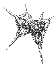
 So I started
sketching them from different angles. When I had the three
dimensional shape in my mind I started to make the final drawing.
I'm afraid I don't use the traditional method of taking exact
measurements. My aim is to make an impression of what I think the
organisms look like.
So I started
sketching them from different angles. When I had the three
dimensional shape in my mind I started to make the final drawing.
I'm afraid I don't use the traditional method of taking exact
measurements. My aim is to make an impression of what I think the
organisms look like.
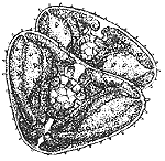
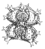
 I made a group
portait of Staurastrum and
related genera. What I found most difficult was showing both the
ornamentation as well as the inner structure. The cell wall is
covered with rows of spikes or knobs. All in a regular pattern.
The inner structure contains two pyrenoids, in each semi-cell.
Between them, in the actual center of the organism I could
sometimes see the nucleus. The chloroplast fills the rest of the
cell. Because of the form of Staurastrum the chloroplast
is split in three parts, each part appears to have two halves. In
the center of the drawing there is a Staurastrum still
attached after cell-division. Some species are not symmetrical,
one half being slightly rotated to the other. After cell division
this results in the two cells lying in the same direction.
I made a group
portait of Staurastrum and
related genera. What I found most difficult was showing both the
ornamentation as well as the inner structure. The cell wall is
covered with rows of spikes or knobs. All in a regular pattern.
The inner structure contains two pyrenoids, in each semi-cell.
Between them, in the actual center of the organism I could
sometimes see the nucleus. The chloroplast fills the rest of the
cell. Because of the form of Staurastrum the chloroplast
is split in three parts, each part appears to have two halves. In
the center of the drawing there is a Staurastrum still
attached after cell-division. Some species are not symmetrical,
one half being slightly rotated to the other. After cell division
this results in the two cells lying in the same direction.
Peridinium
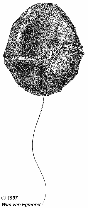 Working with the 100x immersion lens can be a bit messy
but the result is that you can see many details. I examined a
small dinoflagellate found in the same water as the Staurastrum.
When you look very carefully and adjust your microscope as good
as possible you can see the two flagella (one moving through a
groove) and the structure of the plates of the cell wall. The
possibilities of the light microscope are not to be
underestimated. It may be more difficult to see the three
dimensional shape than it is with a electron microscope. But what
we see is alive and we can look right through it.
Working with the 100x immersion lens can be a bit messy
but the result is that you can see many details. I examined a
small dinoflagellate found in the same water as the Staurastrum.
When you look very carefully and adjust your microscope as good
as possible you can see the two flagella (one moving through a
groove) and the structure of the plates of the cell wall. The
possibilities of the light microscope are not to be
underestimated. It may be more difficult to see the three
dimensional shape than it is with a electron microscope. But what
we see is alive and we can look right through it.
My next article will be about three dimensional
photography through an ordinary light microscope and how to make
a stereoscope from a binocular without spending a fortune.
Note: We only used the genus
names. Even with the help of experts there remained several
uncertainties.
The author Wim van Egmond would welcome comments.
Editor's notes:
The desmid illustrations are derived from the original group
portrait. Click here to view a scan of this original drawing.
The Editor and author would like to thank Bill
Ells for useful comments on this article and the desmid
identification.
The images are the copyright of the author, and
should not be either redistributed or used commercially without
seeking the permission of the author via Jan Parmentier.
© Microscopy UK or their
contributors.
Please report any Web problems
or offer general comments to the Micscape Editor,
via the contact on current Micscape Index.
Micscape is the on-line monthly
magazine of the Microscopy UK web
site at Microscopy-UK
WIDTH=1
© Onview.net Ltd, Microscopy-UK, and all contributors 1995 onwards. All rights
reserved. Main site is at www.microscopy-uk.org.uk with full mirror at www.microscopy-uk.net.