|
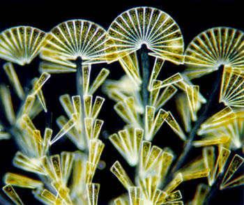
Licmophora in dark field illumination |
by Wim van Egmond
About photographing
epiphytic colonies of marine diatoms
A
mirror of the original article http://www.tip.nl/users/egmond/art_deco.html
on
Wim van Egmond's web site.
| On a quiet October morning in 1997 Micscape's Maurice Smith, Jan Parmentier (chairman of the Dutch microscopy club) and I went for a trip to the Dutch province Zeeland. Zeeland is formed by several peninsulas and has many interesting aspects for microscopists and those who like marine biology. |
|
|
Licmophora in dark field illumination |
| Half the province of Zeeland was flooded in 1953. That's why the dikes were reinforced and a vast system of sluices was build. The most interesting part of these so called deltaworks is it keeps the vulnerable tidal ecosystem intact. We went to one of Jan Parmentier's favourite collecting spots at the Oosterschelde, a little harbour called Burgsluis. The quality of the water of the Oosterschelde is good and the water is very clear. It is fairly easy to get near to the water. Attached to the wooden poles of the pier are all kinds of interesting marine organisms: anemones, several kinds of algae, bryozoans, tunicates, etc. |
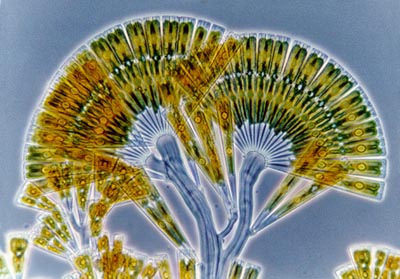 |
The colony in phase contrast illumination. |
| By
laying flat on the pier I could easily reach for them. The real challenge
is then to grab some algae while keeping your belongings inside your pockets.
I filled a little jar with water and selected several species of red algae.
On some of the algae I could see some brown dirt. I knew that dirt growing
on top of something could be very beautiful under the microscope (or just
dirt.) This time I was lucky. No sand grains which make it impossible to
get a flat sample. I found an epiphytic diatom called Licmophora flabellata.
It is a spectacular fan-shaped colony of diatoms.
I tried to make photographs of them. The red algae are rather large compared to the diatoms. When you want to use stronger objectives the sample will be so deep that it is very difficult to get the diatoms focused. So I took a little knife and cut the about 1 mm. sized colony carefully from the red algae. I gently lowered the cover glass and removed water with a tissue until the sample was as flat as possible. Then I could get a sharp picture. I photographed it in phase contrast to show the almost transparent stem. I also took some dark field pictures. |
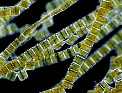 |
The diatom approach to cubism. An epiphytic diatom colony of the genus Rhabdonema. |
| It is very easy to make dark-field illumination yourself. See the article "Making dark-field illumination is easy" to learn how. |
Comments to the author Wim van Egmond are welcomed.
or visit Wim's HOME PAGE
Microscopy
UK Front Page
Micscape
Magazine
Article
Library
all material © Wim van Egmond