When people describe spicules, they generally think of the needle-like structures in sponges which serve both as skeletal support and deterrents to predators. Indeed many spicules are acicular or needle-like; however, a very large number are not. Indeed, a very large volume could be written cataloguing spicules from protists to tunicates. If I had enough time left, I would very much enjoy producing such a work, but I shall have to content myself with producing this very modest, but I hope interesting album.
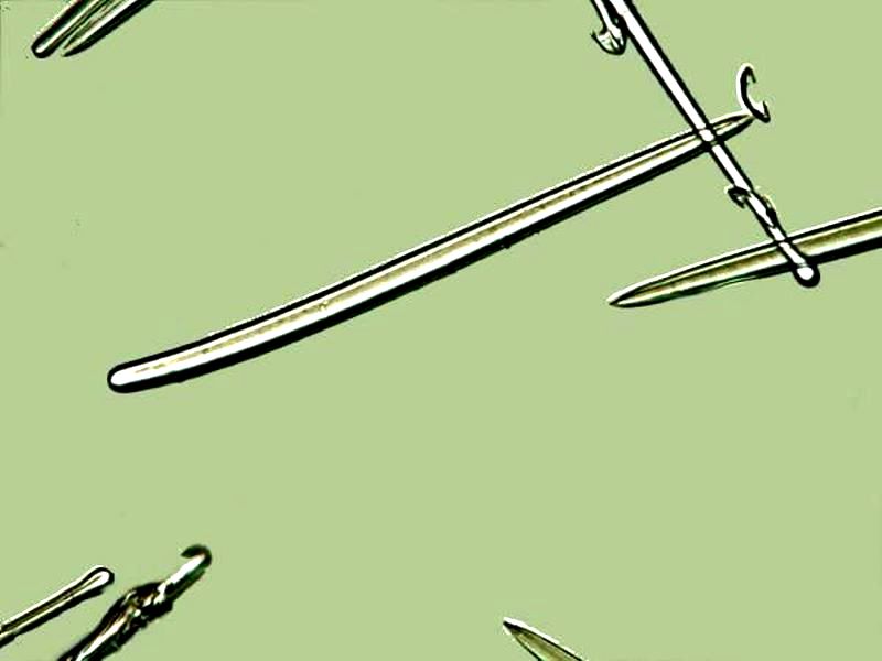
While it’s true that the simple needle-like rod is very common in both calcareous and siliceous forms, it is perhaps the least interesting of spicule forms However, acicular spicules can be the major components of intricate lattices. Here is an example of acicular spicules.
And here is an example of a very complex lattice from a section of the glass sponge, Euplectella aspergillum which is composed of a variety of spicule types, but also a lot of acicular ones.
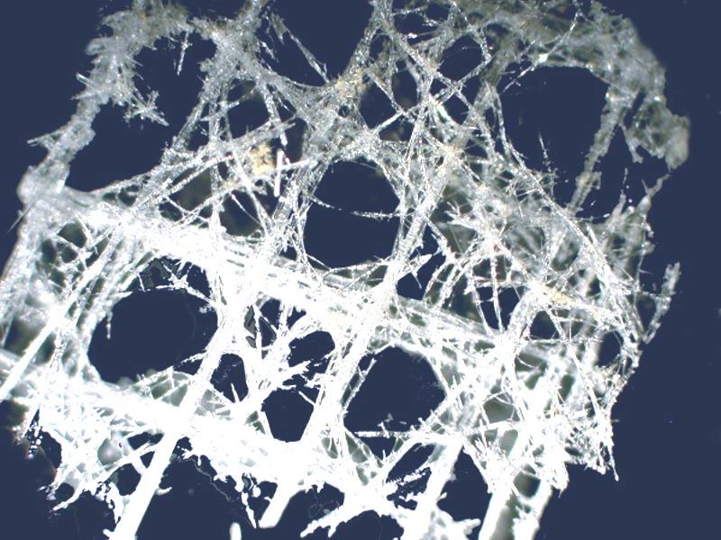
The image when inverted and with the background color slightly altered looks rather like the steel skeletal frame of a building in progress.
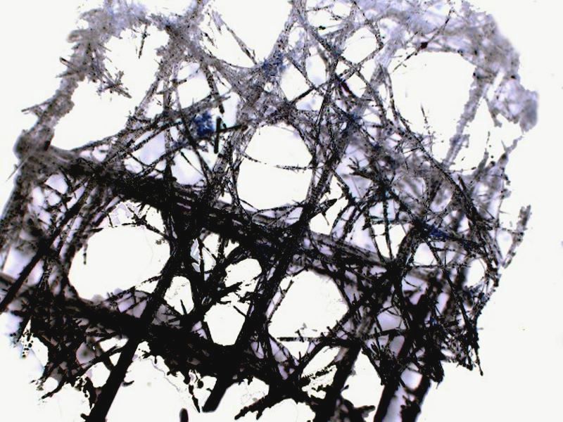
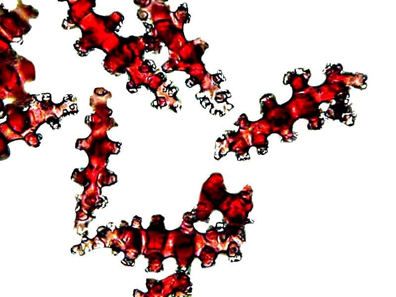
Glass sponges are particularly rich in their different types of spicules as we’ll shall see in a bit. However, first, let’s look at a couple of types of spicules that move beyond the acicular type. The first is from the “sea pansy” (Renilla) which is related to the anemones and “sea pens”. Many soft corals also have this type of spicule which is a rod tapered at each end with tiny knobby structures along the rod.
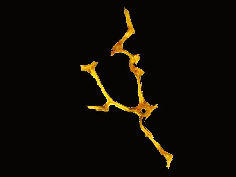
There are some sponges which have a sort of skeletal network which is entirely organic and so might not meet some definitions of spicule. However, it does serve both for support for the sponge and deterrence of predators. Furthermore, one might argue that the difference between a calcareous or siliceous lattice and an organic one is not all that great. In any case, I am including an image of a fragment of such an organic sponge network that rather reminds me of a “goose-stepping” alien.
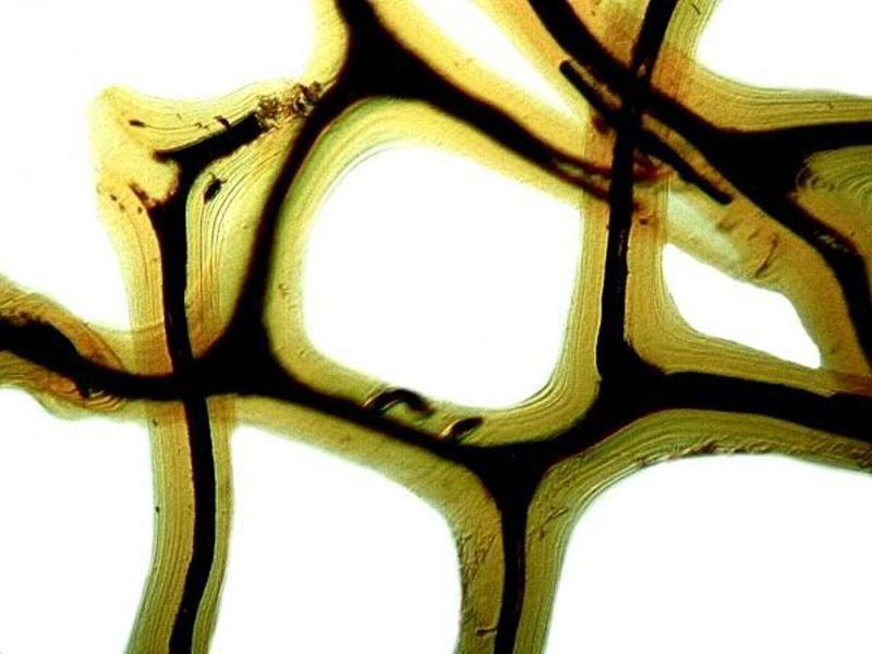
Here is a closeup view of a portion of the network.
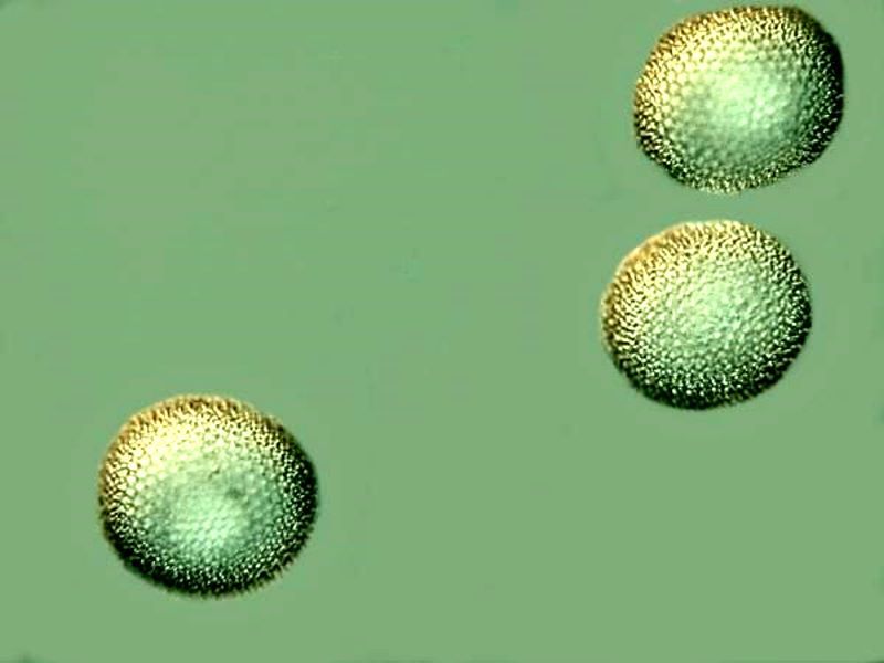
In addition, I came across a coral-like sponge which has some rather surprising spicules; namely, spheres. Here is a view of 3 such spheres using Nomarski DIC. If you look closely, you’ll note that the surfaces are covered with tiny projections.
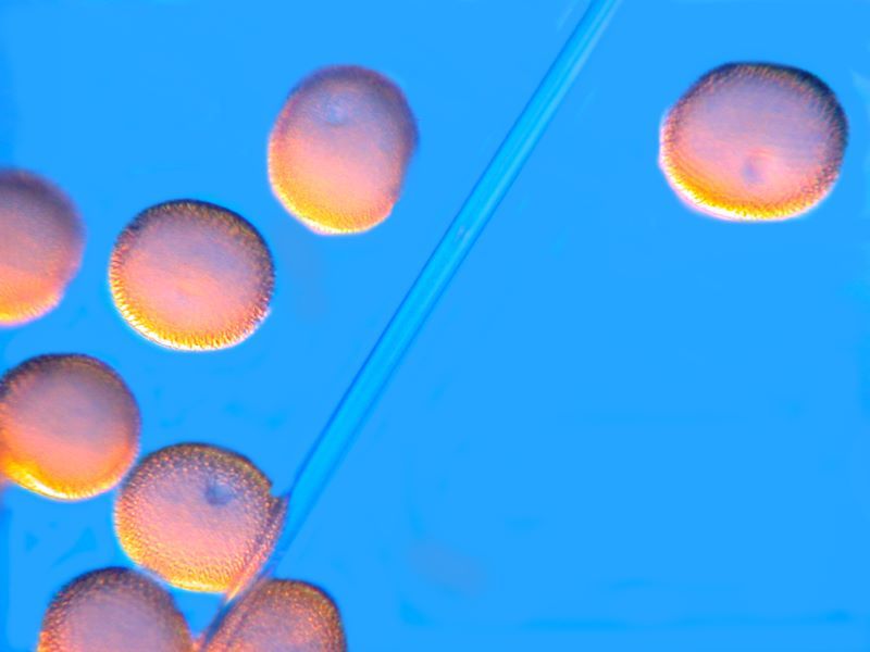
Here’s another view with 8 spheres where I have altered the color contrasts and through the middle of the image you can see the vague outline of an acicular spicule.
However, let us now go to the fertile territory of the spicules in glass sponges. For an overview, I have written several articles on Euplectella and Hyalonema and if you’re interested in digging deeper, you can find them by doing a search on MICSCAPE for Euplectella and/or Hyalonema.
Euplectella is beautifully elegant at the macro-level whereas Hyalonema definitely is not. However, at the microscopic level both reveal remarkably lovely structures.
Let’s begin with a simple example from Hyalonema of what I call the “sword” spicule.
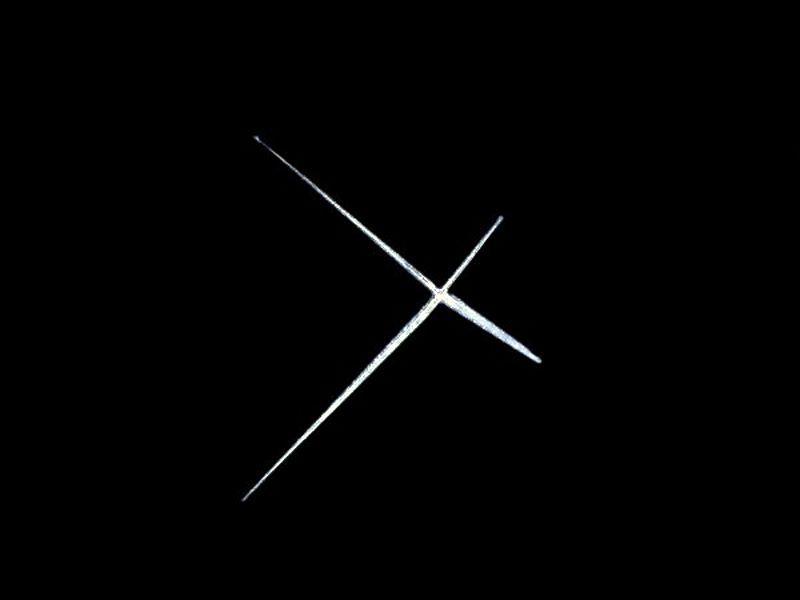
This one would be awkward to wield in battle!
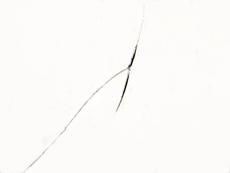
Hyalonema also has spicules of such incredible delicacy that I wonder what possible function they can serve.
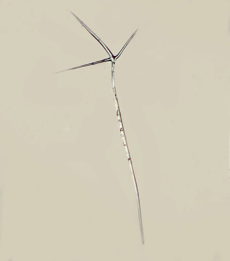
And here is yet another type in the form of tetraxon.
Another remarkable type which I have dubbed the “anchor” spicules, for obvious reasons, is worth a look. I’ll show you 2 views, the first with its long “rope” attachment and the second a closeup of 2 of the anchor tips.
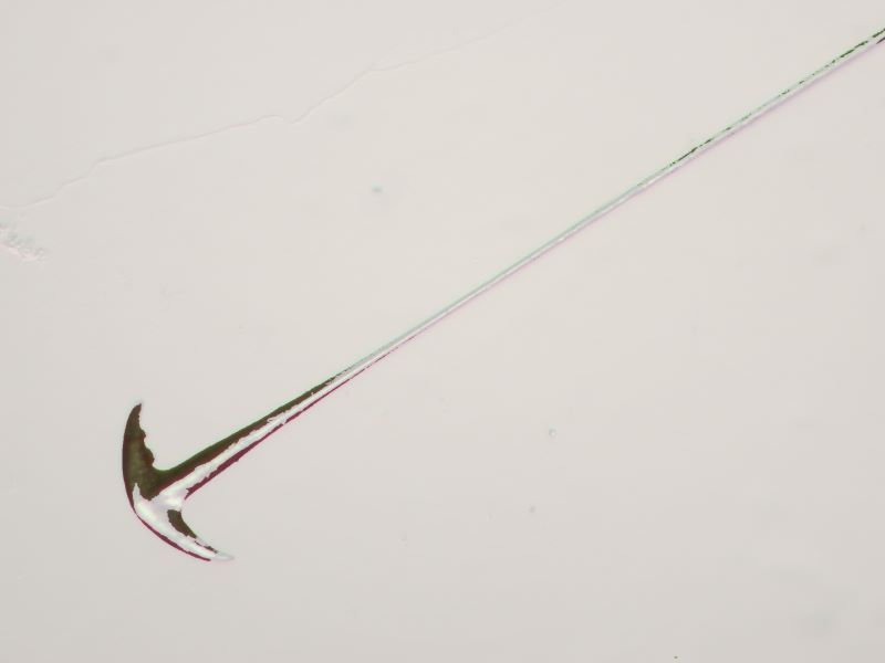
The inverse of this image is positively sinister.
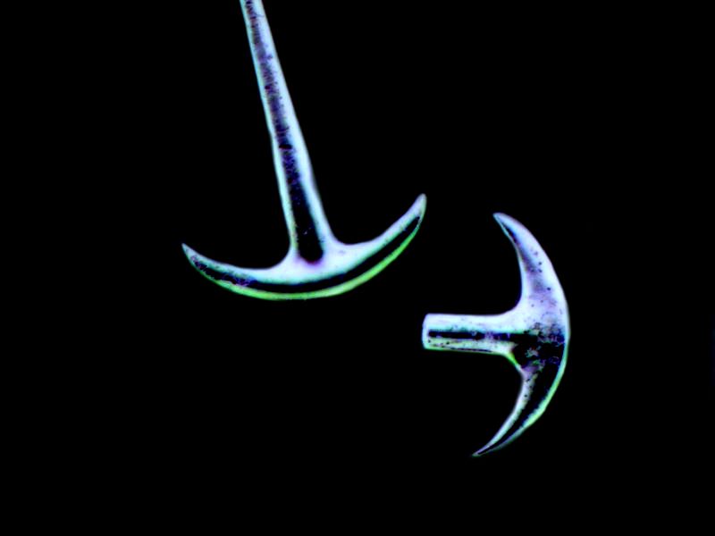
As we’ll see a bit later, sponges are not the only organisms to have anchor-like spicules.
However, Hyalonema is not yet through providing us with surprises and I’ll show you 3 more.
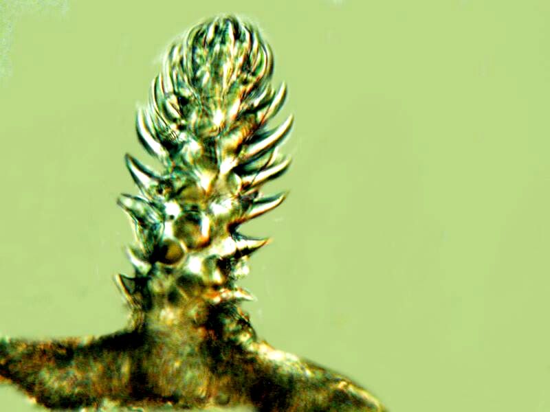
I like to give spicules descriptive names when possible as it helps me keep track of some of the morphological patterns. Below is a cluster of little “tree” spicules.
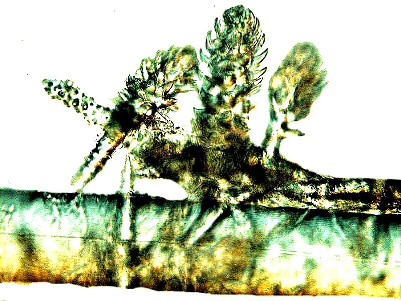
These are actually hexactinellids and sometimes single isolated specimens look like tiny “Christmas trees” on a stand. You can get a hint of that here.
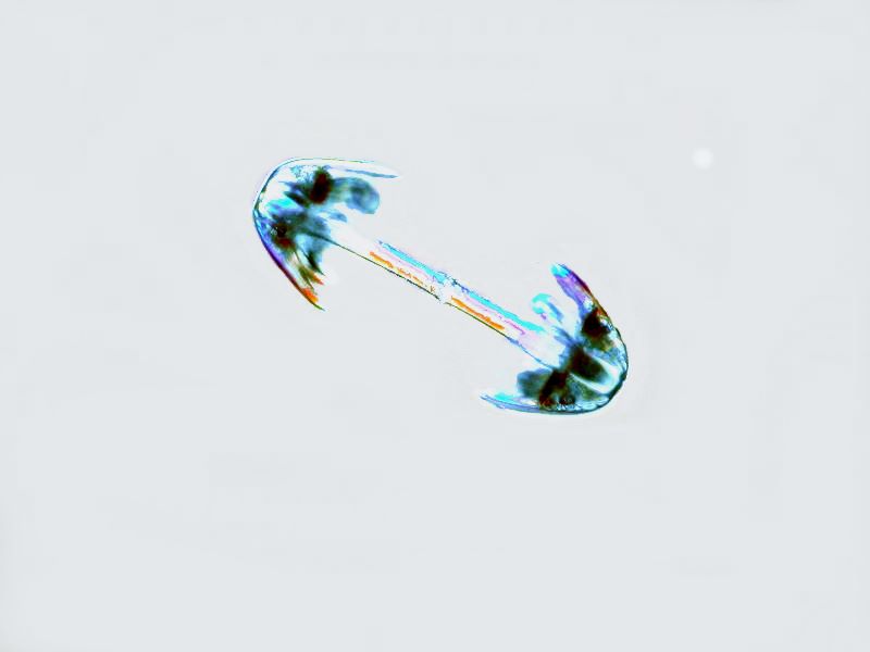
And then, there is the remarkably weird “dumbbell” spicule.
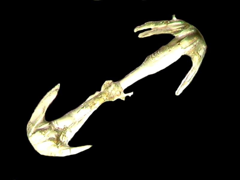
Nature is sometimes a bizarre artist.
The third one makes me think of some variant of a Japanese martial arts throwing star.
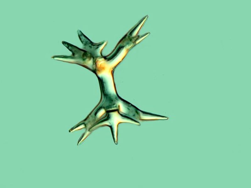
Who would think that all of this morphological strangeness could be packed into one sponge?
Now let’s go back to Euplectella for a couple of moments. Remember this is the Venus Flower Basket with the long “glass tail” of delicate fibers.
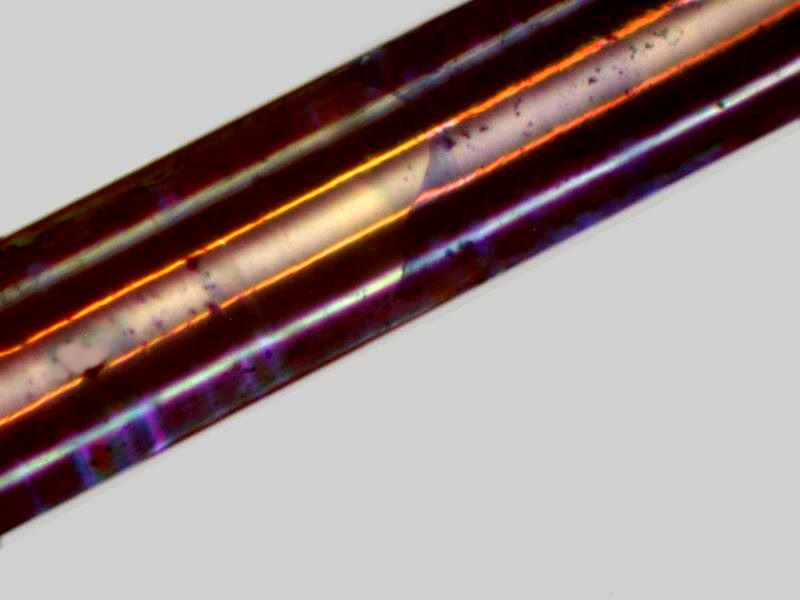
Now, we all know that it’s extremely difficult to stain glass unless you mix dyes into molten glass or forget to wash your room temperature wine glass for a very long time in order to see what the Bordeaux will do to it over the years. Well, I’m rather thick-headed and I was curious about what might happen if I immersed some fragments of Euplectella fibers in some 1% solutions of stains. So, I put some fragments in a half a dozen different such solutions and, frankly, rather forgot about them. When a couple of months later I went back to examine them, I was pleasantly surprised.
I learned from the Toluidine Blue fragments that the fibers were not silky smooth as I had assumed, but ridged, showing the additional material which had been added on; growth rings, if you will.
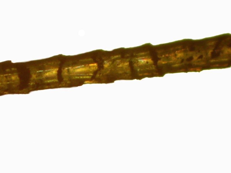
On reflection, this made perfect sense, because one wouldn’t expect fibers over a foot in length to be generated in a single go.
Methylene Blue results suggested that there are fibers within the fiber.
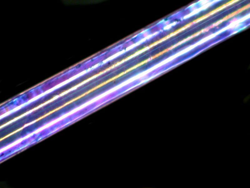
And a similar, although slightly different pattern was obtained with Malachite Green.
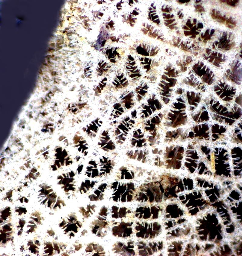
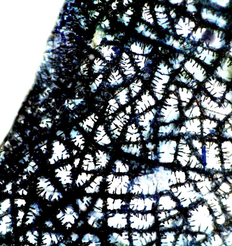
The wonder of all of this is increased when one considers the bewilderingly complex lattices which these spicules fuse to form. Here are 2 views from yet another species of glass sponge. The first images is brightfield, the second uses the application of the invert function.
Now let’s make a major taxonomic leap to a major advanced group of organisms which also display a stunning variety of spicules, namely, the holothuroids or sea cucumbers.
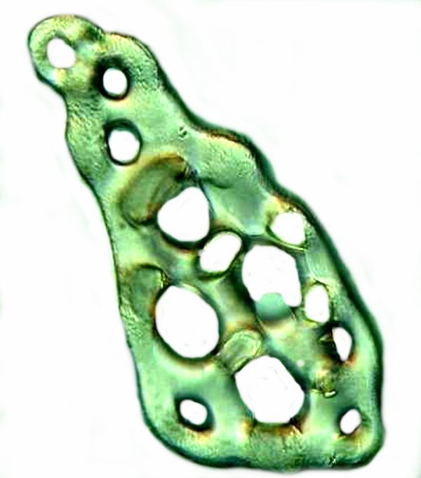
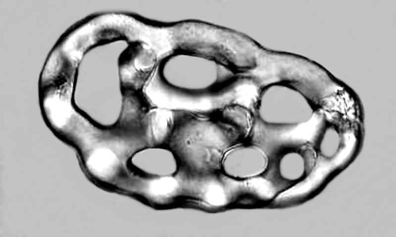
We’ll begin with Eupentacta, a small semi-tropical form which is of interest because the epidermis is densely packed with fenestrated spicules which would make it quite unappetizing to most predators. Most specimens are only 3 or 4 inches in length and packed with thousands of these spicules. I’ll show 2 forms: the first with a color alteration and the other type in monochrome.
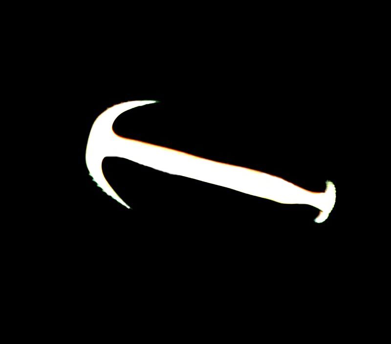
But you ain’t seen nothin’ yet. Remember earlier I told you when we were talking about anchor spicules in sponges that some sea cucumbers have anchors as well. Here is an image of a single one which I isolated.
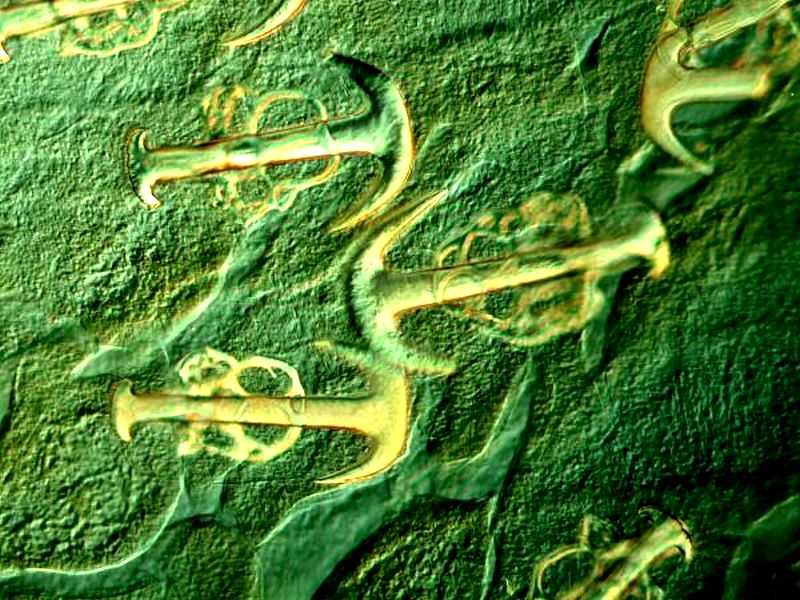
However, a section of tissue showing both the plates and the anchors is much more interesting. Here is a view using Nomarski DIC.
The application of the invert function is helpful in enhancing contrast.
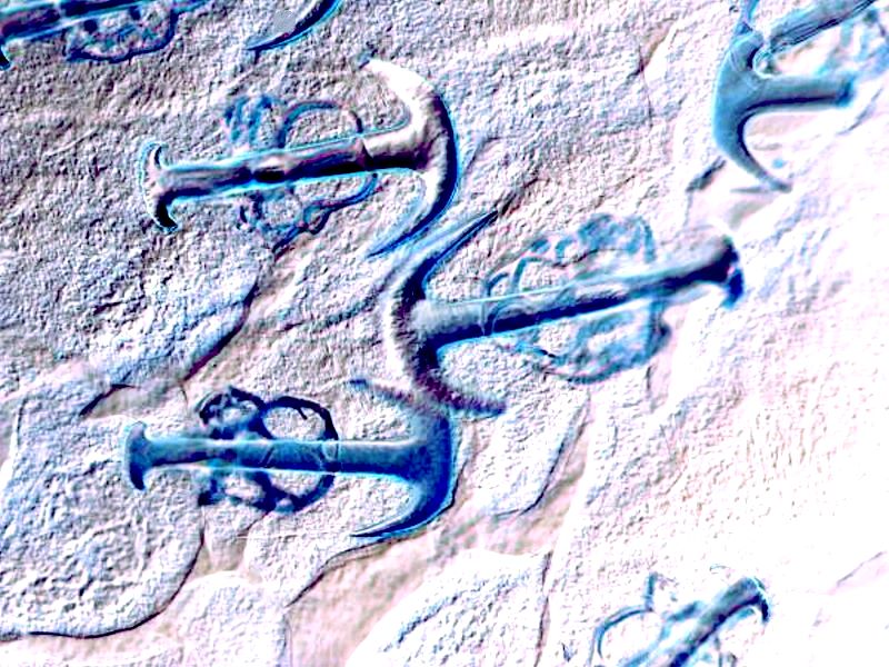
These are from a sea cucumber of the genus Synapta and Victorian mounters quite liked these as subjects.
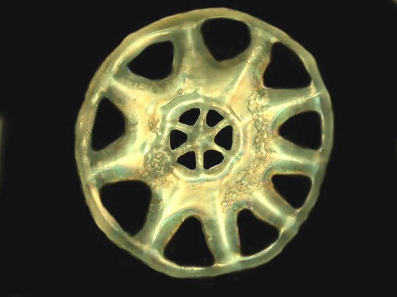
However, even more astonishing are the “wheel” spicules of Chirodota. The sea cucumber itself is rather unprepossessing compared to some of its more flamboyant relatives. It is generally only 3 to 5 inches long, white or light lavender in color with white bumps just under the surface.
It is the bumps that are of extraordinary interest for they are clumps of wheel spicules. So, what does a wheel spicule look like? I isolated a single spicule and here is a closeup view.
Here is another image with 3 wheels photographed using Nomarski DIC.
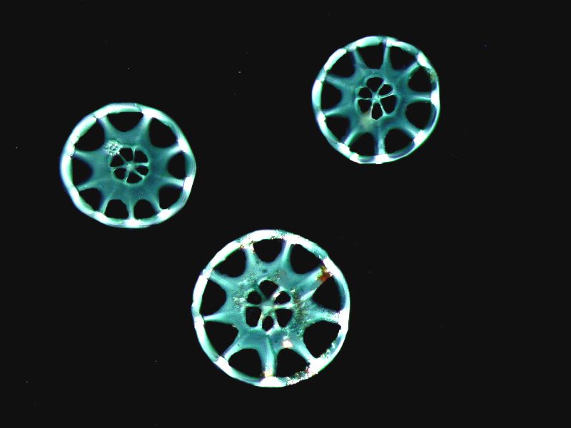
These spicules are from specimens which I obtained from the Philippines. Later however, I managed to obtain a couple of specimens from the Caribbean and immediately began to encounter anomalies which led me into a long and fascinatingly controversial problem about spicule formation in Chirodota. I’ll just show you some examples with brief comments here. If you wish to follow up the problem in detail, you can find the discussion here. Just go to the automated search in MICSCAPE and type in “Howey chirodota” and you’ll find several articles.
First off, let me show you 2 examples of what I regard as incompletely developed spicules. The second one seems further along in the developmental process than the first. Both images are Nomarski DIC.
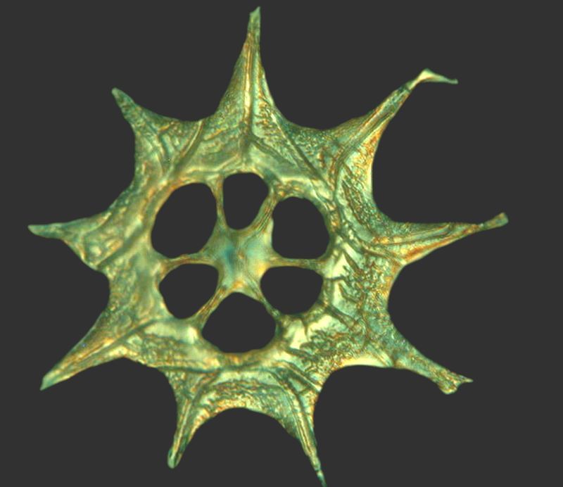
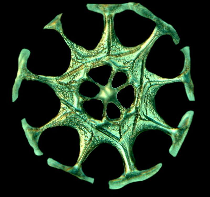
Some have argued against development stages taking the position that these forms are ones either damaged in the process of preparation for observation or from acids in the preservatives which have attacked them. I do not find this at all a convincing view. As I delved deeper, I found a more and more puzzles, such as “baby” wheels alongside “incomplete spicules”. First a Nomarski DIC image and then an inverted color contrast image.
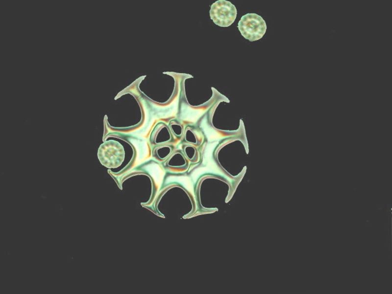
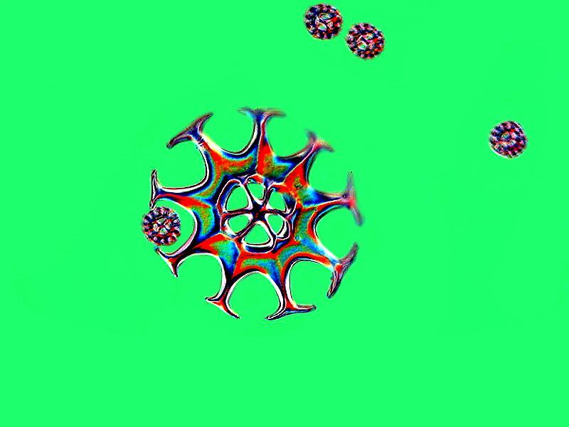
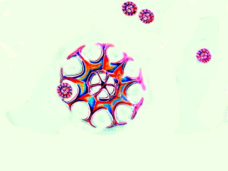
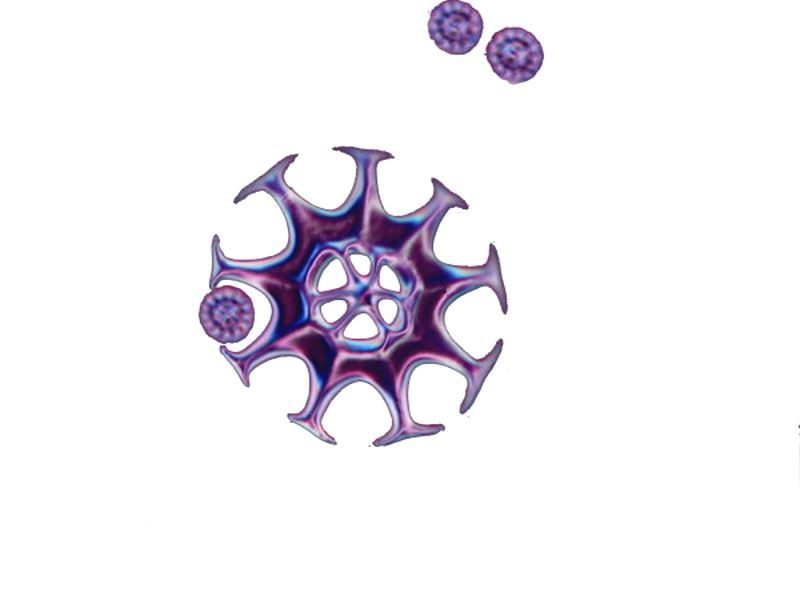
And a view of an “incomplete” form using inverted color contrast. I don’t know how to account for all the variations, but I am quite convinced that there are indeed developmental stages and the processes surrounding them are highly complex. I am especially fond of this image and so I am going to indulge myself and show you 3 more versions as an illustration of what you can do with a bit of computer graphics magic.
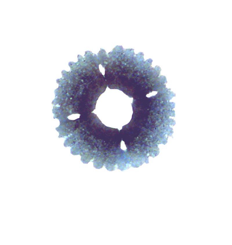
One final marvel extracted from Chirodota. As you know, sea cucumbers have tube feet that they use for locomotion. Some of these feet contain spicules and/or plates as support. Chirodota has at the tip of each tube foot a beautifully constructed circular plate which is itself constructed of several fused sections.
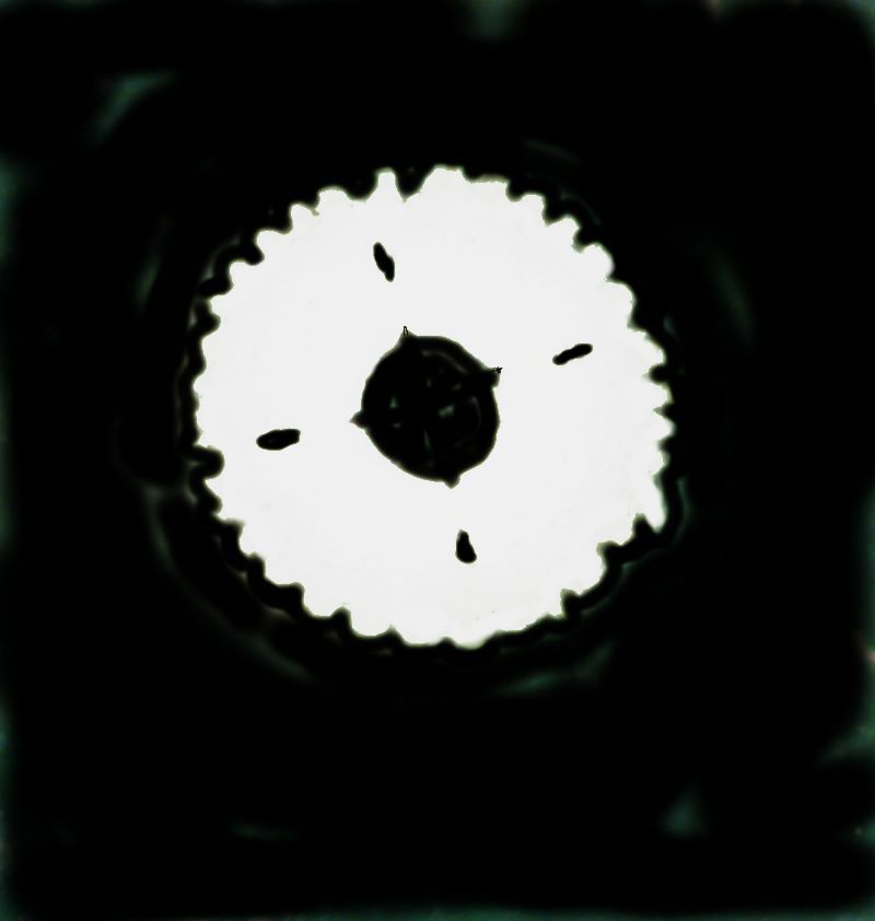
And to conclude, the inverse image of the plate with color contrast.
I hope you have enjoyed this little tour, but even more, I hope that it leads you to look further into the remarkable realm of spicules. There are spicules in some nudibranchs and in some tunicates and I suspect even in some other groups where no one has yet bothered to look. So, start looking; you might be the first to find some in some wonderful, wee organism or, perhaps, even in a relatively large one.
All comments to the author Richard Howey are welcomed.
Editor's note: Visit Richard Howey's new website at http://rhowey.googlepages.com/home where he plans to share aspects of his wide interests.
Microscopy UK Front
Page
Micscape
Magazine
Article
Library
© Microscopy UK or their contributors.
Published in the February 2020 edition of Micscape Magazine.
Please report any Web problems or offer general comments to the Micscape Editor .
Micscape is the on-line monthly magazine of the Microscopy UK website at Microscopy-UK .
©
Onview.net Ltd, Microscopy-UK, and all contributors 1995
onwards. All rights reserved.
Main site is at
www.microscopy-uk.org.uk .