|
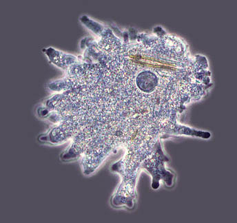
Amoeba proteus in different spot-lights |
Illumination and contrast techniques for the light microscope |
|
Phase contrast |
|
| Phase contrast enables the researcher to see otherwise invisible structures found within the cell body. This technique is invaluable when photographing or filming living cells in action. What the phase contrast system does is to enhance the destructive interference of the light passing through the specimen and medium. | |
|
|
The objective and the condenser both have annuli placed within their respective configuration
so that when these two rings are aligned flagella, cell inclusions and many more biological structures can be seen. The annuli found in the optical train split the waves into two and retard the light waves passing through by a 1/4 th wavelength. The human eye cannot detect these phase differences; the phase contrast microscope enables these out of step wave trains to be brought into focus so the eye can perceive them as changes in amplitude. Depending on the amount of interference will determine the brightness or darkness of the specimen. |
| One disadvantage of the phase contrast system is the halos that are seen around the edges of the specimen. When photographing a specimen in B&W it is best to use a green filter. Most microscope manufacturers make their own phase contrast outfit and they can be quite expensive. | |


Back to An Introduction to Microscopy
Microscopy
UK Front Page
Micscape
Magazine
Article
Library
text & Image © Steve Durr 1999
Web design: Wim van Egmond
© Onview.net Ltd, Microscopy-UK, and all contributors 1995 onwards. All rights reserved. Main site is at www.microscopy-uk.org.uk